. 2014 Dec 17:4:7461.
doi: 10.1038/srep07461.

WP1066 sensitizes oral squamous cell carcinoma cells to cisplatin by targeting STAT3/miR-21 axis

Affiliations

WP1066 sensitizes oral squamous cell carcinoma cells to cisplatin by targeting STAT3/miR-21 axis

Xuan Zhou et al. Sci Rep. .

Abstract

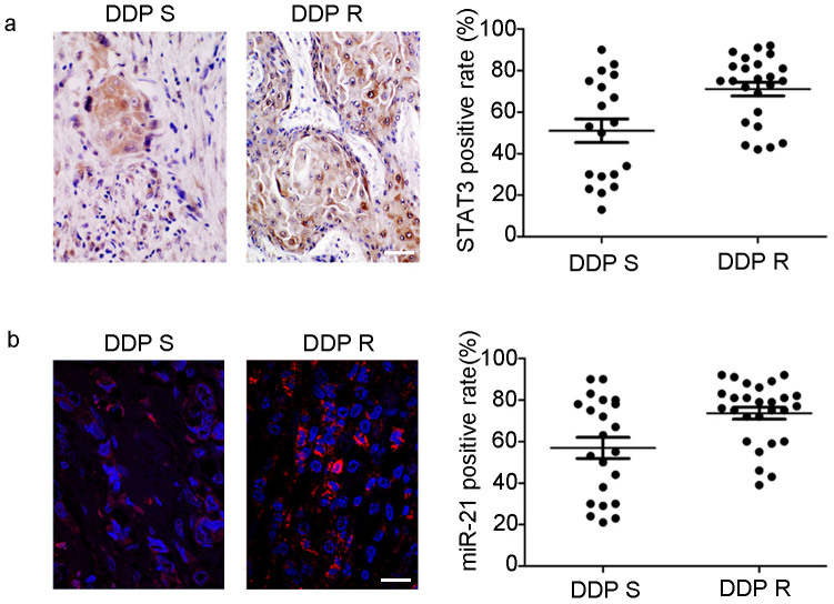
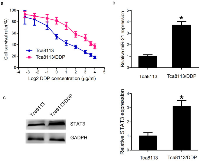
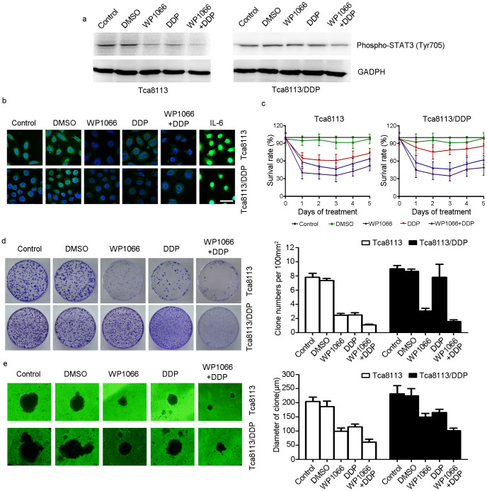
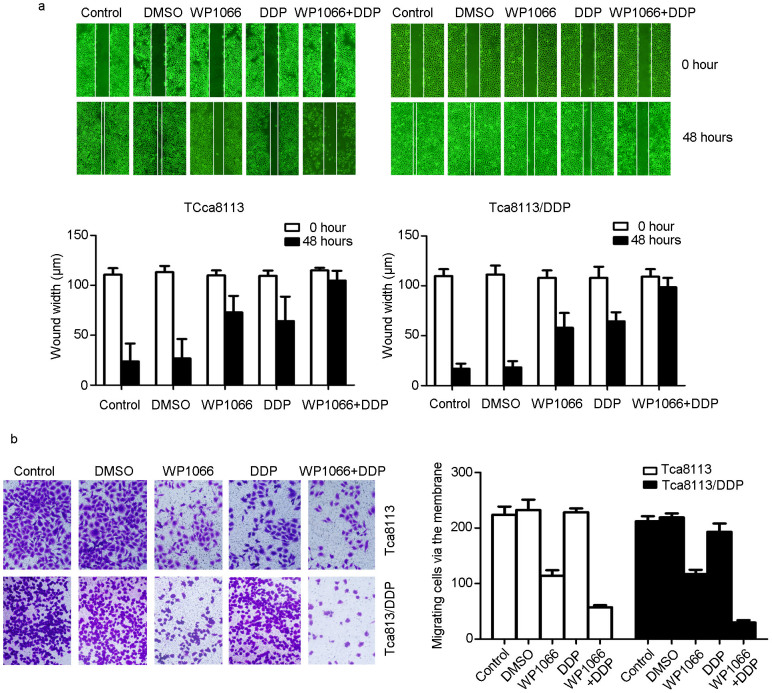
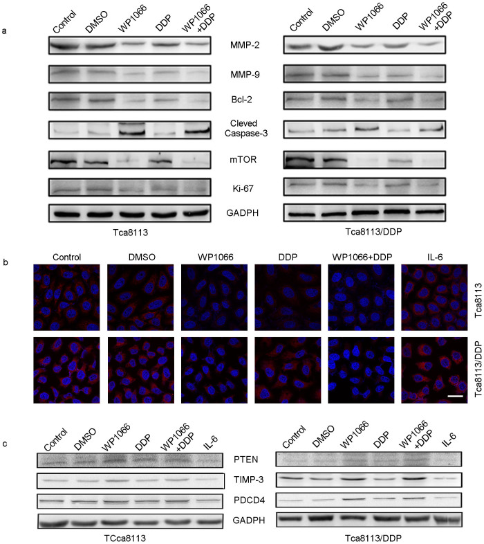
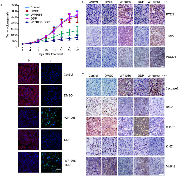
Accumulating evidence reveals that activation of STAT3 and miR-21 contributes to chemoresistance in multiple tumors. We examined the expression of STAT3 and miR-21 in 43 oral squamous cell carcinoma (OSCC) tumors and classified them into cisplatin sensitive or resistant group. Tca8113 and Tca8113/DDP cells were treated with cisplatin (DDP), WP1066 (STAT3 inhibitor) or in combination. MTT, colony formation, wound healing, 3-D culture, and transwell chamber assays were used to evaluate the malignant phenotype of OSCC cells. We evaluated the effect of WP1066 on the expression of STAT3 and miR-21. A Tca8113/DDP OSCC xenograft tumor model was established to evaluate the therapeutic effect of WP1066 in combination with DDP. The expression of STAT3/miR-21 was significantly increased in DDP-resistant OSCC samples and Tca8113/DDP cells compared to its parental cell. Treatment of DDP combined with WP1066 efficiently inhibited Tca8113 and Tca8113/DDP cell proliferation, migration and invasion. STAT3 mediated OSCC cell survival and DDP resistance through upregulating the expression of miR-21 and downregulating miR-21 downstream targets, including PTEN, TIMP3 and PDCD4. WP1066 plus DDP treatment could inhibit Tca8113 and Tca8113/DDP cell growth by inhibiting STAT3 phosphorylation and miR-21 expression. These results indicated that STAT3/miR-21 axis could be a candidate therapeutic target for OSCC chemoresistance.


Figures

Figure 1
Figure 1. STAT3 and miR-21 is over-expressed in DDP resistant OSCC tumor tissues.
(a) In DDP resistant OSCC tumor tissues, STAT3 was highly expressed in the cytoplasm and nucleus of tumor cells than in DDP sensitive OSCC tissues (P < 0.05). (B) In DDP resistant OSCC tissues, ISH staining reveals that miR-21 (red) has a relatively high expression than in DDP sensitive OSCC tissues (P < 0.05) (Bar = 100 μm).
Figure 2
Figure 2. STAT3/miR-21 axis was upregulated in the DDP-resistant Tca8113/DDP cells.
(a) MTT assay indicated that Tca8113/DDP and Tca8113 cells showed different sensitivity to DDP treatment. The cells were incubated in different doses of DDP (0.1, 0.2, 0.5, 1, 2, 4, 8, 12 and 16 μg/ml) for 48 h, and survival rate was determined by MTT assay. (b) QPCR showed that miR-21 was 3.7 folds higher in Tca8113/DDP cells than in Tca8113 cells (P < 0.05). (c) Western blot assay indicated that STAT3 protein expression was 3 folds higher in Tca8113/DDP cells than in Tca8113 cells (P < 0.05). Anti-GAPDH antibody was used as a loading control.
Figure 3
Figure 3. WP1066 sensitized the Tca8113/DDP cells to DDP.
(a) Tca8113 and Tca8113/DDP cells were treated with 5 μM WP1066 and harvested at 48 h. Western blots showed WP1066 and DDP combined treatment inhibited p-STAT3 expression in both cell lines. Anti-GAPDH antibody was used as a loading control. (b) STAT3 was measured by immunofluorescence (IF) method. In WP1066 alone and DDP+ WP1066 treated group, STAT3 expression was downregulated significantly in both Tca8113 and Tca8113/DDP cells. In IL-6 treated cell, STAT3 expression was upregulated (Bar = 20 μm). (c) MTT assay indicated that WP1066 and DDP combined treatment inhibited both Tca8113 and Tca8113/DDP cell lines growth in vitro. (d) Clone formation assay indicated that Tca8113/DDP cells were resistant to DDP. DDP + WP1066 treatment inhibited Tca8113/DDP and Tca8113 cell growth effectively. (e) 3-D culture assay showed that DDP + WP1066 treatment significantly inhibited Tca8113/DDP and Tca8113 cell growth on martrigel matrix.
Figure 4
Figure 4. WP1066 sensitized OSCC cells to DDP and reduced migration capability.
Scratch (Wound healing) and transwell assays were used to assess cell migration and invasion capability at 48 h after treatment. (a) Scratch assay has shown that combination of WP1066 and DDP could delay the scratch healing in Tca8113 and Tca8113/DDP cells. (b) Transwell assay has shown that combination of WP1066 and DDP could decrease the invasion of Tca8113 and Tca8113/DDP cells.
Figure 5
Figure 5. WP1066 combined DDP could inhibit the expression of miR-21 and its target genes.
ISH was used to determine the expression of miR-21 in treated OSCC cells; and Western blot was used to detect the expression of its target genes. (a) In Tca8113 and Tca8113/DDP cells, WP1066 combined with DDP treatment could inhibit MMP2/9, Bcl-2, mTOR, Ki67 expression, while in DDP + WP1066 treated group, Caspase-3 expression was upregulated. (b) In Tca8113 and Tca8113/DDP cells, WP1066 combined with DDP treatment could induce the expression of miR-21 (Red signal labeled by Cy3, cell nuclei was labeled by DAPI). IL-6 treatment could inhibit the expression of miR-21. (Bar = 20 μm) (c) In Tca8113 and Tca8113/DDP cells, DDP + WP1066 treatment could induce the expression of PTEN, TIMP3 and PDCD4. While IL-6 treatment could inhibit their expression in both cell lines. The gels were cropped, but those gels were run under the same experimental condition.
Figure 6
Figure 6. Combination of WP1066 and DDP inhibited Tca8113/DDP xenograft tumor growth.
(a) The growth curves of blank control, DMSO, WP1066-treated, DDP, and DDP + WP1066 treated Tca8113/DDP xenograft tumors. The growth rate in WP1066, DDP and DDP + WP1066 group were inhibited compared to control and DMSO groups. (b) ISH showed that, in WP1006, WP1066 and DDP treated Tca8113/DDP tumors, miR-21 expression was inhibited (Red signal labeled by Cy3, cell nuclei was labeled by DAPI). (c) TUNEL assay showed an induced apoptotic nucleus (green) in WP1066 combined DDP-treated Tca8113/DDP tumors. (d) IHC staining showed the upregulation of PTEN, TIMP-3, and PDCD4 in WP1066-treated and DDP + WP1066 treated group.(e) IHC assay was used to determine the expression of tumor growth related proteins in Tca8113/DDP xenograft tumors. Downregulation of mTOR, Ki67, Bcl-2, and MMP-2 was observed in WP1066 and WP1066 combined DDP-treated group. In combined treated group Caspase-3 expression was upregulated. (Bar = 100 μm).

References

    1. Pulte D. & Brenner H. Changes in survival in head and neck cancers in the late 20th and early 21st century: a period analysis. Oncologist. 15, 994–1001 (2010). - PMC - PubMed
    1. Lam L., Logan R. M. & Luke C. Epidemiological analysis of tongue cancer in South Australia for the 24-year period, 1977-2001. Aust. Dent. J. 51, 16–22 (2006). - PubMed
    1. Cullen K. J. et al. Glutathione S-transferase pi amplification is associated with cisplatin resistance in head and neck squamous cell carcinoma cell lines and primary tumors. Cancer Res. 63, 8097–8102 (2003). - PubMed
    1. Jacobs C. et al. A phase III randomized study comparing cisplatin and fluorouracil as single agents and in combination for advanced squamous cell carcinoma of the head and neck. J Clin Oncol. 10, 257–263 (1992). - PubMed
    1. Shen D. W., Pouliot L. M., Hall M. D. & Gottesman M. M. Cisplatin resistance: a cellular self-defense mechanism resulting from multiple epigenetic and genetic changes. Pharmacol Rev. 64, 706–721 (2012). - PMC - PubMed

Publication types

MeSH terms