. 2014 Dec 17:14:310.
doi: 10.1186/s12866-014-0310-7.

Plasminogen activation by staphylokinase enhances local spreading of S. aureus in skin infections

Affiliations

Plasminogen activation by staphylokinase enhances local spreading of S. aureus in skin infections

Marijke Peetermans et al. BMC Microbiol. .

Abstract

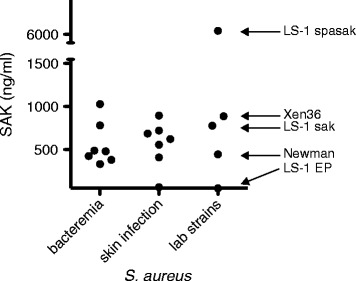
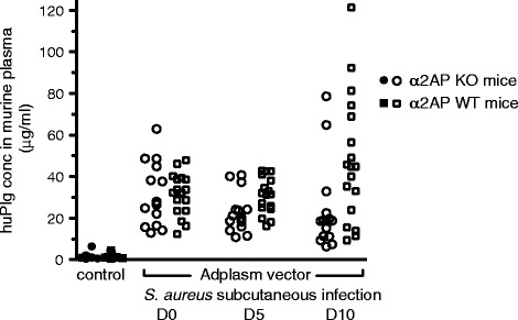
Background: Staphylococcus aureus (S. aureus) is a frequent cause of skin and soft tissue infections. A unique feature of S. aureus is the combined presence of coagulases that trigger fibrin formation and of the plasminogen activator staphylokinase (SAK). Whereas the importance of fibrin generation for S. aureus virulence has been established, the role of SAK remains unclear. We studied the role of plasminogen activation by SAK in a skin infection model in mice and evaluated the impact of alpha-2-antiplasmin (α2AP) deficiency on the spreading and proteolytic activity of S. aureus skin infections. The species-selectivity of SAK was overcome by adenoviral expression of human plasminogen. Bacterial spread and density was assessed non-invasively by imaging the bioluminescence of S. aureus Xen36.

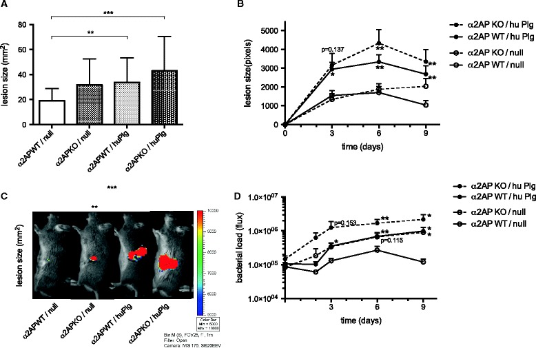
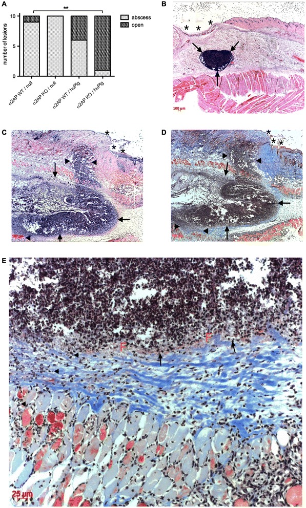
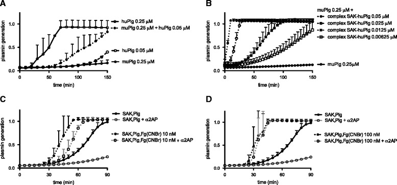
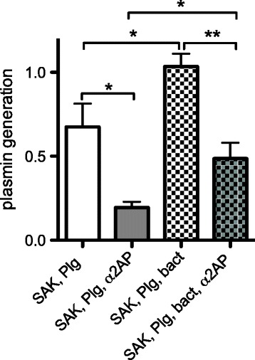
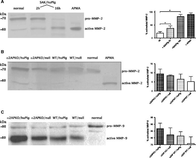
Results: SAK-mediated plasmin activity increased the local invasiveness of S. aureus, leading to larger lesions with skin disruption as well as decreased bacterial clearance by the host. Even though fibrin and bacterial surfaces protected SAK-mediated plasmin activity from inhibition by α2AP, the deficiency of α2AP resulted in increased bacterial spreading. SAK-mediated plasmin also induced secondary activation of gelatinases, shown both in vitro and in lesions from the in vivo model.

Conclusion: SAK contributes to the phenotype of S. aureus skin infections by enhancing bacterial spreading as a result of fibrinolytic and proteolytic activation.


Figures

Figure 1
Figure 1
Bacterial staphylokinase production. Staphylokinase (SAK) production of different S. aureus strains after overnight culture, as assessed by ELISA. SAK secretion of the bioluminescent strain S. aureus Xen36 is comparable to relevant clinical S. aureus strains from bacteremia with cutaneous origin and from skin infection. SAK production of reference lab strains, including a SAK-negative (LS-1 EP) and a SAK-overproducing (LS-1 spasak) S. aureus strain are included as controls.
Figure 2
Figure 2
Adenoviral-mediated human plasminogen expression. Human plasminogen (huPlg) expression in murine plasma after administration of 5 × 1010 viral particles of adenoviral vector Adplasm. Day 0 is the day of subcutaneous infection with S. aureus Xen36, 7 to 11 days after adenoviral injection. Values from Adnull injected mice are included as negative controls. The expression of huPlg during the whole course of the subcutaneous infection experiment allows the selective interaction of staphylokinase with huPlg.
Figure 3
Figure 3
SAK-mediated plasmin activity increased infectious skin lesion size and bacterial load. A. Macroscopically apparent lesion size 10 days after subcutaneous inoculation with S. aureus Xen36. Mean and SD for skin lesions in WT/null mice (n = 12), α2AP KO/null mice (n = 11), WT/huPlg mice (n = 17) and α2AP KO/huPlg mice (n = 14), respectively. B. Lesion size: S. aureus Xen36 possesses a stable copy of the modified Photorhabdus luminescens luxABCDE operon. Evolution of bacterial spread was assessed non-invasively by bioluminescence image analysis of the surface area with signal > threshold. Mean and SEM for lesion size in the 4 groups. Dimensions for α2AP KO/huPlg mice and WT/huPlg mice are compared to WT/null mice. C. Examples of bioluminescence photographs of the left flank lesion for representative animals of the 4 groups at day 9. D. Bacterial load: signal intensity of the infectious lesion site, in photons/s through a defined region of interest, which was used for all lesions. Bacterial density analysis shows that bacterial clearance is hampered by enhanced proteolytic activity. Mean and SEM for signal intensity in the 4 groups. Bacterial loads for α2AP KO/huPlg, α2AP KO/null and WT/huPlg mice are compared to WT/null mice. *denotes P < 0.05, **P < 0.01, ***P < 0.001.
Figure 4
Figure 4
Infectious skin lesions at day 3 after subcutaneous infection with S. aureus Xen36. A. Macroscopic aspect of day 3 lesions. More open lesions with skin rupture are observed in α2AP KO/huPlg mice, compared to WT/null mice (P <0.01). B. Hematoxyllin-eosin staining of lesional skin from WT/null mouse showing a small abscess collection (arrows) without disruption of overlying skin (*). C. Hematoxyllin-eosin staining of lesional skin from α2AP KO/huPlg mouse showing a large, less well-defined abscess collection (arrows), with extension (arrowheads) from the initial infection site both towards the overlying skin (*) (with resulting skin disruption and crust formation) and towards the underlying subdermal tissue and muscularis. D-E. Martius Scarlet Blue staining of the same lesion reveals a zone of fibrin deposition (F, red) at the periphery of the initial abscess site (arrows), but the infection has spread past this border of fibrin, through collagen fibers (blue), into underlying tissue layers (arrowheads).
Figure 5
Figure 5
Species-selectivity and fibrin-specificity of staphylokinase. A. Species-selectivity of staphylokinase (SAK) for human plasminogen (huPlg). Plasmin generation by adding SAK (6.25 nM) to either huPlg (0.25 μM), murine plasminogen (muPlg 0.25 μM), or a mixture of muPlg (0.25 μM) with huPlg (0.05 μM or 20% of the muPlg concentration, comparable to the level of huPlg in murine plasma after adenoviral-mediated huPlg expression). Plasmin generation was quantitated by conversion of the chromogenic substrate S-2403 and assessed in a microtiter plate ELISA reader at 405 nm. Mean and SD from 3 independent experiments. B. Plasmin generation by a preformed equimolar mixture of SAK with huPlg, added to muPlg (0.25 μM), highlighting that low levels of huPlg in a background of muPlg can induce efficient SAK-dependent plasmin generation. Mean and SD from 3 independent experiments. C-D. The complex of SAK with human plasmin (SAK-huPli) is protected from inhibition by alpha-2-antiplasmin (α2AP) in the presence of fibrin analogues. Plasmin generation by SAK (6.25 nM) in a mixture of muPlg (0.25 μM) and huPlg (0.05 μM); with or without α2AP (0.125 μM) and either in the absence or presence of Fg(CNBr) (C. and D. for Fg(CNBr) 10 nM and 100 nM, respectively). Mean and SD from 3 independent experiments.
Figure 6
Figure 6
SAK-huPli complex is protected from inhibition by α 2 AP in the presence of S. aureus bacterial surfaces. S. aureus bacterial surfaces enhance SAK-mediated plasmin generation and partially protect the SAK-huPli complex from inhibition by α2AP. Plasmin generation by SAK (6.25 nM) in a mixture of murine (0.25 μM) and human (0.05 μM) plasminogen, with or without α2AP (0.125 μM) and in the absence or presence of washed SAK-negative S. aureus LS-1 EP (OD600 2.0, 15% vol/vol). Plasmin generation was quantitated by conversion of the chromogenic substrate S-2403 and assessed in a microtiter plate ELISA reader at 405 nm. Mean and SD from 3 independent experiments. *denotes P < 0.05, **P < 0.01.
Figure 7
Figure 7
Gelatinase activation by SAK in skin tissue. A. Gelatin zymogram showing pro-MMP-2 and active MMP-2 in murine skin and subcutaneous tissue protein extract after incubation with staphylokinase (SAK) and human plasminogen (huPlg). Activation with APMA, a chemical MMP-activator, was used as a positive control. Quantitation of MMP-2 activation (active/total), data are mean ± SD from 3 independent experiments. *denotes P < 0.05. B. Gelatin zymogram showing pro-MMP-2 and active MMP-2 in day 3 lesional skin samples. A sample from normal skin and an APMA-activated normal skin extract are included as controls. Quantitation of MMP-2 activation (active/total), data are mean ± SD from 3 independent experiments. P for trend <0.05. C. MMP-9 activation in day 3 lesional skin samples, assessed by Western blot. A sample from normal skin is included as control. Quantitation of MMP-9 activation (active/total), data are mean ± SD from 3 independent experiments. P for trend <0.05.

References

    1. Ray GT, Suaya JA, Baxter R. Microbiology of skin and soft tissue infections in the age of community-acquired methicillin-resistant Staphylococcus aureus. Diagn Microbiol Infect Dis. 2013;76:24–30. doi: 10.1016/j.diagmicrobio.2013.02.020. - DOI - PubMed
    1. Sader HS, Farrell DJ, Jones RN. Antimicrobial susceptibility of Gram-positive cocci isolated from skin and skin-structure infections in European medical centres. Int J Antimicrob Agents. 2010;36:28–32. doi: 10.1016/j.ijantimicag.2010.03.016. - DOI - PubMed
    1. Vanassche T, Verhaegen J, Peetermans WE, Van Ryn J, Cheng A, Schneewind O, Hoylaerts MF, Verhamme P. Inhibition of staphylothrombin by dabigatran reduces Staphylococcus aureus virulence. J Thromb Haemost. 2011;9:2436–2446. doi: 10.1111/j.1538-7836.2011.04529.x. - DOI - PubMed
    1. Vanassche T, Peetermans M, Van Aelst L, Peetermans WE, Verhaegen J, Missiakas D, Schneewind O, Hoylaerts M, Verhamme P. The role of staphylothrombin-mediated fibrin deposition in catheter-related Staphylococcus aureus infections. J Infect Dis. 2013;208:92–100. doi: 10.1093/infdis/jit130. - DOI - PMC - PubMed
    1. McAdow M, Kim HK, Dedent AC, Hendrickx AP, Schneewind O, Missiakas DM. Preventing Staphylococcus aureus sepsis through the inhibition of its agglutination in blood. PLoS Pathog. 2011;7:e1002307. doi: 10.1371/journal.ppat.1002307. - DOI - PMC - PubMed

Publication types

MeSH terms