. 2015 Mar 31;35(4):543-53.
doi: 10.1038/jcbfm.2014.227.

Premyelinated central axons express neurotoxic NMDA receptors: relevance to early developing white-matter injury

Affiliations

Premyelinated central axons express neurotoxic NMDA receptors: relevance to early developing white-matter injury

Tahani Huria et al. J Cereb Blood Flow Metab. .

Abstract

Ischemic-type injury to developing white matter is associated with the significant clinical condition cerebral palsy and with the cognitive deficits associated with premature birth. Premyelinated axons are the major cellular component of fetal white matter and loss of axon function underlies the disability, but the cellular mechanisms producing ischemic injury to premyelinated axons have not previously been described. Injury was found to require longer periods of modelled ischemia than at latter developmental points. Ischemia produced initial hyperexcitability in axons followed by loss of function after Na(+) and Ca(2+) influx. N-methyl-D-aspartate- (NMDA) type glutamate receptor (GluR) agonists potentiated axon injury while antagonists were protective. The NMDA GluR obligatory Nr1 subunit colocalized with markers of small premyelinated axons and expression was found at focal regions of axon injury. Ischemic injury of glial cells present in early developing white matter was NMDA GluR independent. Axons in human postconception week 18 to 23 white matter had a uniform prediameter expansion phenotype and postembedded immuno-gold labelling showed Nr1 subunit expression on the membrane of these axons, demonstrating a shared key neuropathologic feature with the rodent model. Premyelinated central axons therefore express high levels of functional NMDA GluRs that confer sensitivity to ischemic injury.


Figures

Figure 1
Figure 1
Ischemic injury in premyelinated central white matter. (A) Three representative plots of compound action potential (CAP) amplitude in P2 rat optic nerves (RONs) exposed to 90 minutes of oxygen–glucose deprivation (OGD). Note the variable initial rise in CAP amplitude followed by a gradual decline which frequently did not reach zero, followed by the general absence in CAP recovery after reintroduction of artificial cerebrospinal fluid (aCSF). Typical CAP recorded before and after 90 minutes OGD is shown in the inset. (B) Mean data showing the effects of 90 minutes OGD perfusion on CAP amplitude. (C) The effects of Ca2+ removal from the perfusate. Note that under control conditions (filled symbols), zero-Ca2+ (indicated by the top bar) evokes a transient CAP decline. In the test experiment (open symbols), OGD has no additional effect than that produce by Ca2+ removal alone. (D) Data summary showing significantly reduced CAP loss in P2 RON after 90 minutes OGD under either zero-Ca2+ or zero-Na+ conditions (***P<0.001; error bars are s.e.m.).
Figure 2
Figure 2
The significance of excitotoxicity. (A) N-methyl-D-aspartate (NMDA) glutamate receptor (GluR) activation (coperfusion with 1 mmol/L NMDA and 10 μmol/L glycine) for 90 minutes evoked a limited delayed decline in the compound action potential (CAP) under normoxic conditions. (B, C) NMDA receptor block with 1 μmol/L MK-801 or 1 μmol/L memantine increased recovery from oxygen–glucose deprivation (OGD). (D) Data summary showing the effects of NMDA receptor agonists and antagonists on the injury produced by OGD (***P<0.001 versus artificial cerebrospinal fluid (aCSF) of OGD as appropriate).
Figure 3
Figure 3
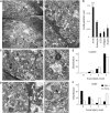
Ultrastructural colocalization of Nr1 on P2 axons correlates with injury. (A) Immunolocalization of Nr1 protein in P2 rat optic nerve (RON) (all ultra-micrographs are cross-sections). Left: Axons surround a glial process (‘gp'), boxed area shown at high magnification below. Typical axon features are microtubules, vesicular-tubular inclusions, diameter <0.4 μm, and the absence of glial ensheathment. Three gold-particles (arrows) are localized to the axolemma of two neighboring axons. Right: Axons proximal and distal to a glial process have gold particles aligned with the axolemma. (B) Blind quantification of gold-particle density in axons, astrocytes, extracellular space (ECS), peri-neural collagen, and glial nuclei. The data show significantly higher staining density in axons versus all other groups (**P<0.01; n=7 nerve sections). (C) Nr1 immunolocalization after 90 minutes oxygen–glucose deprivation (OGD)/60 minutes recovery. Axon injury is variable with some axons showing axoplasmic swelling and/or flocculent debris, swollen mitochondria and microtubule loss. Gold particles are localized to the axolemma of badly damaged axon, boxed area shown at high magnification to the right. (D) Similar images showing two gold particles localized to the axolemma of a damaged axon. (E) Blinded viability scoring of control, showing little pathology and no difference between Nr1(+) axons and Nr1(−) axons (n=400 axons analyzed from 8 sections). (F) A similar analysis after OGD/recovery. Note that viability scores are shifted to lower values for both Nr1(+) axons and Nr1(−) axons, but this effect is greatest in Nr1(+) axons (n=398 axons analyzed).
Figure 4
Figure 4
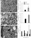
N-methyl-D-aspartate (NMDA) receptor block is protective against oxygen–glucose deprivation (OGD)-induced disruption of axon structure. (AC) Representative ultramicrographs from control (A), post 90 minutes OGD+60 minutes recovery (B) and postOGD performed in the presence of 1 μmol/L MK-801 (C). (D) Blinded counting of axon density in the three groups shows significantly lower number of identifiable axons after OGD (n=254 axons, 6 sections) compared with control perfused rat optic nerves (RONs) (n=213 axons from 5 sections), an effect that was significantly reduced by NMDA receptor block (n=576 axons from 7 sections). (E) Blinded axon viability scores were significantly lower in identifiable axons after OGD (n=398 axons from 6 sections), compared with RONs continually perfused with aCSF (n=298 axons from 6 sections), an affect that was prevented by NMDA receptor block (n=327 axons from 6 sections). (F) The distribution of axon viability scores in these three conditions, showing that NMDA receptor block restores a more normal pattern of viability. Scale bar=0.5 μm. ‘*'=P<0.05 versus control; ‘**'=P<0.01 versus control; ‘***'=P<0.001 versus control; §§=P<0.01 versus OGD; §§§=P<0.001 versus OGD.
Figure 5
Figure 5
N-methyl-D-aspartate (NMDA) receptors do not mediate Ca2+ influx or cell death in P2 rat optic nerve (RON) astrocytes. (A) FURA-2-AM loaded astrocytes in P2 RON at various times after the onset of oxygen–glucose deprivation (OGD) in the presence of 1 μmol/L MK-801 (arrow indicates cell shown in B). (B) Changes in 340/380 ratio corresponding to [Ca2+]i in a representative cell (open squares, top data set, left-hand scale). Note the elevation in 340/380 ratio after the onset of OGD in the presence of MK-801. The 360 intensity of this cell is also plotted (filled circles, lower data, right hand scale). Note the collapse in 360 intensity (arrow), indicative of cell lysis. (C) The extent of cell death in P2 RON astrocytes under control conditions (n=198 cells from 6 nerves), during 90 minutes of OGD (n=173 cells from 6 nerves) and during 90 minutes of OGD in the presence of MK-801 (n=192 cells from 6 nerves). Note the significant rise in cell death during OGD compared with control, and the absence of protection in the presence of MK-801 (***P<0.001 versus control). (D) Histograms showing the times when cell death occurred under the three conditions. (E) GFAP staining (red) and FURA-2 localization (green) in fixed, FURA-2-AM loaded P2 RON. Images overlaid to the right, showing that all FURA-2 filled cells are GFAP(+). Scale=1 μm.
Figure 6
Figure 6
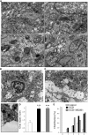
Glial injury is non-NMDA glutamate receptor (GluR)-R mediated. (A) Typical P2 rat optic nerve (RON) astrocyte subjected to 90 minutes OGD/60 minutes recovery shown at low magnification (Top, left) with the three boxed areas at high gain below and to the right. Typical features include glial filaments (‘GF'), dark bodies (‘db'), and glycogen particles. Golgi apparatus (‘ga'), endoplasmic reticulum (‘er'), and mitochondria (‘Mt') appear intact but swollen. Cell membrane integrity has been lost in this cell (arrows, bottom right panel). (B) Dead astrocyte after oxygen–glucose deprivation (OGD)/recovery+1 μmol/L MK-801. The cell has lost membrane integrity and organelles are in continuity with the expanded extracellular space. (C) Nerve perimeter after this protocol, showing swollen and severely disrupted astrocyte processes (‘*') of the glial limitans adjacent to the basement membrane (arrows); boxed area at high magnification below left, showing intact axons with normal mitochondria and microtubules neighboring the grossly swollen and fragmented glial processes. (D) Cell injury scoring for control, 90 minutes OGD/60 minutes recovery and following the same protocol in the presence of 1 μmol/L MK-801. Astrocytes show high levels of injury after OGD, and effect that was not reduced by MK-801 (n=22 astrocytes in each group, ***P<0.001 versus control). (E) The distribution pattern of injury scores under these three conditions. NMDA, N-methyl-D-aspartate.
Figure 7
Figure 7
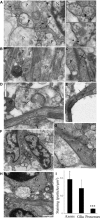
Nr1 expression in mid-gestation human cortex. (A) Cortical postconception week (PCW) 22 gray matter is rich in small diameter axons (e.g., ‘*') surrounding larger cell processes (‘P') and neuron soma (‘N'). Note the characteristic microtubules within axons. Regions of intense reactivity (arrows) are evident within axolemma (boxed areas at high gain to the right), in addition to staining within the neuron and processes. Scale=1 μm. (BG) Subcortical intermediate zone. (B) Typical axonal staining of small diameter axons mainly in long section, and cell processes that may be glial. Scale=500 nm. (C) A localized region of intense Nr1 reactivity within the axoplasm underlying a region of axon extension, where the axolemma protrudes from the axon (longitudinally oriented microtubules identify the axon). Scale=500 nm. (D) Axolemma gold staining at the site of glial contact (note axonal microtubules). Scale=500 nm. (E) Gold staining within the axoplasm of an axon, Scale=500 nm. (F) Staining within the cytoplasm of two glial cells (‘A'). Cell identification is difficult but the nuclear morphology suggests they are astroglial. (G) An astrocyte process identified by glial filaments (e.g., arrow heads) is Nr1(+) (arrows). Scale=500 nm. (H) Reactivity within the cytoplasm and cell membrane of two neighboring glioblasts. Scale=1 μm. (I) Gold particle density in positively identified axons, glia somata, and cell processes in the intermediate zone. Counts were collected from 12 sections, ‘***'=P<0.001 versus axon density.

Similar articles

Cited by

References

    1. Schmitz Y, Luccarelli J, Kim M, Wang M, Sulzer D. Glutamate controls growth rate and branching of dopaminergic axons. J Neurosci. 2009;29:11973–11981. - PMC - PubMed
    1. Ehlers MD, Fung ET, O'Brien RJ, Huganir RL. Splice variant-specific interaction of the NMDA receptor subunit NR1 with neuronal intermediate filaments. J Neurosci. 1998;18:720–730. - PMC - PubMed
    1. Herkert M, Rottger S, Becker CM. The NMDA receptor subunit NR2B of neonatal rat brain: complex formation and enrichment in axonal growth cones. Eur J Neurosci. 1998;10:1553–1562. - PubMed
    1. Wang PY, Petralia RS, Wang YX, Wenthold RJ, Brenowitz SD. Functional NMDA receptors at axonal growth cones of young hippocampal neurons. J Neurosci. 2011;31:9289–9297. - PMC - PubMed
    1. Aoki C, Venkatesan C, Go CG, Mong JA, Dawson TM. Cellular and subcellular localization of NMDA-R1 subunit immunoreactivity in the visual cortex of adult and neonatal rats. J Neurosci. 1994;14:5202–5222. - PMC - PubMed

Publication types

Substances