. 2014 Dec 17:11:212.
doi: 10.1186/s12974-014-0212-5.

Inhibition of the NLRP3 inflammasome provides neuroprotection in rats following amygdala kindling-induced status epilepticus

Affiliations

Inhibition of the NLRP3 inflammasome provides neuroprotection in rats following amygdala kindling-induced status epilepticus

Xiang-Fei Meng et al. J Neuroinflammation. .

Abstract

Background: NLRP3 inflammasome is proposed to regulate inflammation in several neurological diseases, but its role in epilepsy remains largely unknown. This study aimed to investigate the role of the NLRP3 inflammasome in neuroinflammation, spontaneous recurrent seizures (SRS) and hippocampal neuronal loss in rat brain following amygdala kindling-induced status epilepticus (SE).

Methods: We detected the protein levels of IL-1β and NLRP3 inflammasome components by Western blot in the hippocampus of shams and SE rats at different time points following SE. To further examine whether the activation of the NLRP3 inflammasome contributes to SE-associated neuronal damage, we employed a nonviral strategy to knock down NLRP3 and caspase-1 expression in brain before undergoing SE. Proinflammatory cytokine levels and hippocampal neuronal loss were evaluated at 12 hours and at 6 weeks following SE respectively in these NLRP3 and caspase-1 deficient rats. Meanwhile, SRS occurrence was evaluated through a 4-week video recording started 2 weeks after SE in these NLRP3 and caspase-1 deficient rats.

Results: IL-1β levels and NLRP3 inflammasome components levels dramatically increased at 3 hours after SE, and reached a maximum at 12 hours after SE compared with the control group. Knock down of NLRP3 or caspase-1 decreased the levels of IL-1β and IL-18 at 12 hours after SE, which was accompanied by a significant suppression in the development and severity of SRS during the chronic epileptic phase. Meanwhile, knock down of NLRP3 or caspase-1 led to a remarkable reduction of hippocampal neuronal loss in the CA1 and CA3 area of the hippocampus at 6 weeks after SE.

Conclusions: Our study provides the first evidence that the NLRP3 inflammasome was significantly up-regulated following SE. More importantly, we show that inhibition of the NLRP3 inflammasome provides neuroprotection in rats following SE. These findings suggest that NLRP3 may represent a potential target for the treatment of epileptogenesis.


Figures
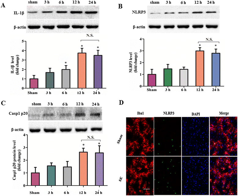
Figure 1
Figure 1
Expression profiles of the nucleotide binding and oligomerization domain-like receptor family pyrin domain-containing 3 (NLRP3) inflammasome components following status epilepticus (SE). (A-C) Western blot assay for the expression profiles of cleaved IL-1β (18 kDa) (A), NLRP3 (106 kDa) (B) and cleaved caspase-1 (20 kDa) (C) in the hippocampus of sham rat at 12 hours following the sham operation of amygdala stimulation (without any electrical stimulation) and SE rat at 3, 6, 12, and 24 hours following amygdala stimulation. Data are expressed as a fold change relative to sham group. n = 6 rat per group and per time point. Error bars represent mean ± standard deviation. *P < 0.05 versus sham control group. NS: not significant versus 12 hours following SE. (D) Representative photographs of immunofluorescence staining for NLRP3 (green) expression in microglia (Iba-1, red) in the hippocampal area 12 hours following SE. Scale bars: 100 μm.
Figure 2
Figure 2
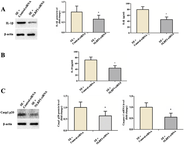
Downregulation of nucleotide binding and oligomerization domain-like receptor family pyrin domain-containing 3 (NLRP3) by siRNA led to significant reduction of proinflammatory cytokines and cleaved caspase-1. (A) The expression level of cleaved IL-1β (18 kDa) was detected by Western blot analysis and ELISA. β-actin was used as loading control. (B) The expression level of cleaved IL-18 was detected by ELISA. (C) The protein expression level of active caspase-1 (20 kDa) was analyzed using the Western blot assay. The gene expression level of caspase-1 was detected by quantitative real-time PCR. Data are expressed as a fold change relative to SE rat infused with control siRNA. All data are shown as mean ± standard deviation (n = 6 per group). *P < 0.05 versus control siRNA treatment.
Figure 3
Figure 3
Downregulation of nucleotide binding and oligomerization domain-like receptor family pyrin domain-containing 3 (NLRP3) by siRNA attenuated hippocampal neuronal loss in status epilepticus (SE rat). (A) Representative photo of Nissl-staining in CA1 region and CA3 region of the rat hippocampus. Neurons with intact morphology were identified as surviving neurons. Scale bars: 50 μm. The neuronal survival rate was defined as follows: Neuronal surviving rate (%) = 100 × (Count of surviving neurons/Total count of neurons). (B) Neuronal death was detected using the TUNEL staining in the hippocampus of sham rats and SE rats. Photos were converted to black and white to obtain a better contrast ratio. Neurons with deep black nuclei were identified as TUNEL-positive neurons (indicated by red arrows). Scale bars: 50 μm. The percentage of TUNEL-positive neurons was defined as follows: 100 × (Count of TUNEL-positive neurons/Total count of neurons). Columns represent mean ± standard deviation (n = 6 per group). *P < 0.05 versus control siRNA treatment. TUNEL, terminal deoxynucleotidyl transferase-mediated dUTP end-labeling.
Figure 4
Figure 4
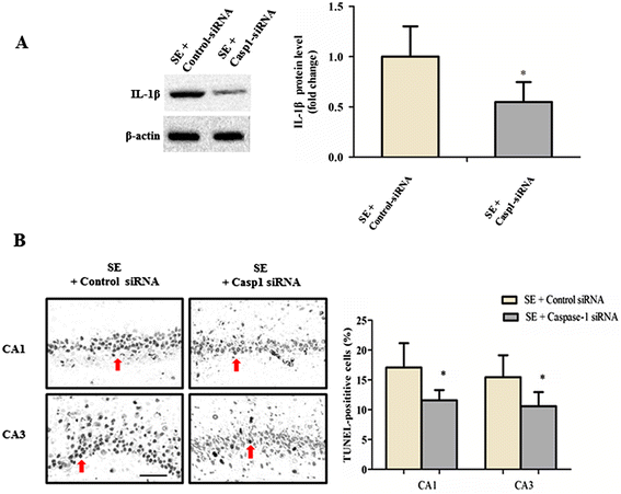
Caspase-1 inhibition ameliorated neuroinflammation and hippocampal neuronal loss in status epilepticus (SE) rat. (A) The expression level of cleaved IL-1β (18 kDa) was detected by Western blot. β-actin was used as loading control. Data are expressed as a fold change relative to SE rat infused with control siRNA. *P < 0.05 versus control siRNA treated group. (B) Neuronal death was detected using the TUNEL staining in the CA1 and CA3 region of the hippocampus. Photos were converted to black and white to obtain a better contrast ratio. Neurons with deep black nuclei were identified as TUNEL-positive neurons (indicated by red arrows). Scale bars: 50 μm. The percentage of TUNEL-positive neurons was defined as follows: 100× (Count of TUNEL-positive neurons/Total count of neurons). All data are shown as mean ± standard deviation (n = 6 per group). *P < 0.05 versus control siRNA-treated group. TUNEL, terminal deoxynucleotidyl transferase-mediated dUTP end-labeling.
Figure 5
Figure 5
A schematic linking the nucleotide binding and oligomerization domain-like receptor family pyrin domain-containing 3 (NLRP3) inflammasome activation to status epilepticus (SE) pathogenesis. Activation of the NLRP3 inflammasome typically requires a bimodal signaling pathway. A Toll-like receptor (TLR)-dependent priming step activates the NF-κB-dependent transcription of NLRP3 and the pro-forms of the proinflammatory cytokines (which are IL-1β and IL-18). NLRP3-activating stimulation agents provide a second signal in the form of K+ efflux, cytosolic release of mitochondria-derived factors such as reactive oxygen species (ROS), cardiolipin, and oxidized mitochondrial DNA (mtDNA). Note that acidic extracellular pH represents a novel stimulation agent for triggering NLRP3 inflammasome activation. Oligomerization of NLRP3 is followed by recruitment of the adaptor molecule apoptosis-associated speck-like protein containing a caspase recruitment domain (ASC) and the pro-form of caspase-1, leading to the activation (cleavage) of caspase-1. Activated caspase-1 in turn catalyzes the cleavage of IL-1β and IL-18. This event may lead to changes in brain parenchyma such as leakage of the blood-brain barrier (BBB), neuronal hyperexcitability and excitotoxicity as well as neuronal damage which contribute to lowering the threshold for seizure induction and thus to trigger epileptogenesis. Activation of innate immune mechanisms during epileptogenesis can recruit inflammatory cells from the periphery which perpetuate inflammation, thus activating a vicious cycle that in turn fosters aberrant hyperexcitability. The onset of SE can in turn further promote inflammation via the production of proinflammatory cytokines.

References

    1. Gastaut H. Clinical and electroencephalographical classification of epileptic seizures. Epilepsia. 1970;11:102–113. doi: 10.1111/j.1528-1157.1970.tb03871.x. - DOI - PubMed
    1. Vezzani A, French J, Bartfai T, Baram TZ. The role of inflammation in epilepsy. Nat Rev Neurol. 2011;7:31–40. doi: 10.1038/nrneurol.2010.178. - DOI - PMC - PubMed
    1. Dube C, Vezzani A, Behrens M, Bartfai T, Baram TZ. Interleukin-1beta contributes to the generation of experimental febrile seizures. Ann Neurol. 2005;57:152–155. doi: 10.1002/ana.20358. - DOI - PMC - PubMed
    1. Vezzani A, Friedman A, Dingledine RJ. The role of inflammation in epileptogenesis. Neuropharmacology. 2013;69:16–24. doi: 10.1016/j.neuropharm.2012.04.004. - DOI - PMC - PubMed
    1. Vezzani A, Baram TZ. New roles for interleukin-1 Beta in the mechanisms of epilepsy. Epilepsy Curr. 2007;7:45–50. doi: 10.1111/j.1535-7511.2007.00165.x. - DOI - PMC - PubMed

Publication types

MeSH terms

LinkOut - more resources