. 2015 Mar;24(3):341-53.
doi: 10.1002/pro.2621. Epub 2015 Jan 13.

Exploring the folding pathway of green fluorescent protein through disulfide engineering

Affiliations

Exploring the folding pathway of green fluorescent protein through disulfide engineering

Derek J Pitman et al. Protein Sci. 2015 Mar.

Abstract

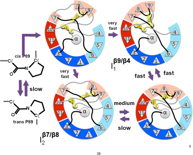
We have introduced two disulfide crosslinks into the loop regions on opposite ends of the beta barrel in superfolder green fluorescent protein (GFP) in order to better understand the nature of its folding pathway. When the disulfide on the side opposite the N/C-termini is formed, folding is 2× faster, unfolding is 2000× slower, and the protein is stabilized by 16 kJ/mol. But when the disulfide bond on the side of the termini is formed we see little change in the kinetics and stability. The stabilization upon combining the two crosslinks is approximately additive. When the kinetic effects are broken down into multiple phases, we observe Hammond behavior in the upward shift of the kinetic m-value of unfolding. We use these results in conjunction with structural analysis to assign folding intermediates to two parallel folding pathways. The data are consistent with a view that the two fastest transition states of folding are "barrel closing" steps. The slower of the two phases passes through an intermediate with the barrel opening occurring between strands 7 and 8, while the faster phase opens between 9 and 4. We conclude that disulfide crosslink-induced perturbations in kinetics are useful for mapping the protein folding pathway.

Keywords: folding pathway; kinetics; leave-one-out GFP; protein design.


Figures

Figure 1
Figure 1
A model derived from the crystal structure of superfolder GFP (PDB:2B3P) showing the locations of the disulfide mutations. The backbone is shown in cartoon, with beta strand 7 shown in red and beta strand 8 shown in green. The modeled disulfides are shown as colored spheres. The A disulfide is highlighted with a red box, and the B disulfide is highlighted with a black box. The close-ups of the highlights show the local environment of the modeled disulfides in stereo.
Figure 2
Figure 2
Fluorescence spectra for the disulfide mutants. Excitation scans were from 300 to 495 nm, and emission scans were from 500 to 600 nm. Scans were performed for all three mutants: the AB disulfide (red), the single A disulfide (green) and the single B disulfide (blue) under oxidizing (solid lines) and reducing (dashed lines) conditions. Fluorescence was normalized by protein concentration and plotted relative to the maximum signal of OPT-GFP under the same conditions (black). Under oxidizing conditions, the mutants show about 40–50% of the fluorescence signal of OPT-GFP; under reducing conditions, this number is 60–70%.
Figure 3
Figure 3
Plot of relative amplitudes and rate constants of refolding for the three mutants. This plot shows the kinetic refolding parameters for the AB mutant (black), the single A disulfide mutant (white) and the single B disulfide mutant (gray). The reduced state is plotted with circles, and the oxidized state is plotted with squares. The rate axis is on a logarithmic scale, and the error bars denote ± 1 S.D. in both directions.
Figure 4
Figure 4
Effects of disulfide bonds on the folding energy landscape. F, folded state; ‡, transition state; I, intermediate states 1 (red) and 2 (blue); U, unfolded state. Red: fast phase. Blue: medium phase. Red arrows indicate changes relative to the reduced state. (a) Native (reduced) landscape. (b) Landscape with the A disulfide present. Folded state is more stable and unfolding is slower. The transition states for both fast and medium phases are shifted towards the unfolded state as shown by the higher m-value. (c) Landscape with the B disulfide present. The medium phase is unaffected by the presence of the disulfide. The fast phase transition state is shifted towards the unfolded state and is faster. (d) Landscape with both A and B present. Both folding and unfolding are slower for both phases and both transition states are shifted towards the unfolded state.
Figure 5
Figure 5
Proposed barrel-closing intermediates. The fast phase intermediate I1 has a break in the barrel between strands 9 and 4. The medium phase intermediate I2 has a break in the barrel between strands 7 and 8. An unstable intermediate may exist in which both of the barrel breaks are present. The slow phase is the isomerization of the backbone at proline 89 producing an off-pathway intermediate in the unfolded state.

References

    1. Jones LN, Simon M, Watts NR, Booy FP, Steven AC, Parry DAD. Intermediate filament structure: hard α-keratin. Biophys Chem. 1997;68:83–93. - PubMed
    1. Anfinsen CB. Principles that govern the folding of protein chains. Science. 1973;181:223–230. - PubMed
    1. Ruoppolo M, Vinci F, Klink TA, Raines RT, Marino G. Contribution of individual disulfide bonds to the oxidative folding of Ribonuclease A. Biochemistry. 2000;39:12033–12042. - PubMed
    1. Wilkinson B, Gilbert HF. Protein disulfide isomerase. Biochim Biophys Acta, Proteins Proteomics. 2004;1699:35–44. - PubMed
    1. Holmgren A. Thioredoxin and glutaredoxin systems. J Biol Chem. 1989;264:13963–13966. - PubMed

Publication types

Associated data

LinkOut - more resources