Observational Study
. 2015 Mar;70(3):219-28.
doi: 10.1136/thoraxjnl-2014-206225. Epub 2014 Dec 16.

Increased expression of GDF-15 may mediate ICU-acquired weakness by down-regulating muscle microRNAs

Affiliations
Observational Study

Increased expression of GDF-15 may mediate ICU-acquired weakness by down-regulating muscle microRNAs

S A A Bloch et al. Thorax. 2015 Mar.

Abstract

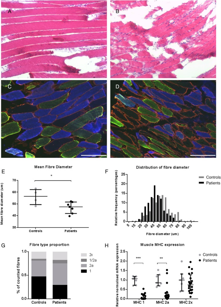
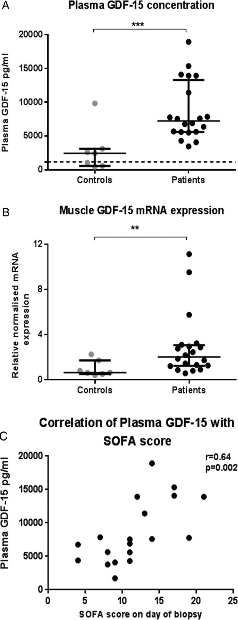
Rationale: The molecular mechanisms underlying the muscle atrophy of intensive care unit-acquired weakness (ICUAW) are poorly understood. We hypothesised that increased circulating and muscle growth and differentiation factor-15 (GDF-15) causes atrophy in ICUAW by changing expression of key microRNAs.

Objectives: To investigate GDF-15 and microRNA expression in patients with ICUAW and to elucidate possible mechanisms by which they cause muscle atrophy in vivo and in vitro.

Methods: In an observational study, 20 patients with ICUAW and seven elective surgical patients (controls) underwent rectus femoris muscle biopsy and blood sampling. mRNA and microRNA expression of target genes were examined in muscle specimens and GDF-15 protein concentration quantified in plasma. The effects of GDF-15 on C2C12 myotubes in vitro were examined.

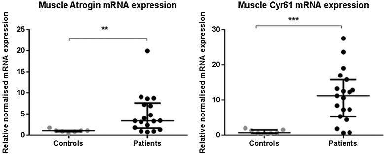
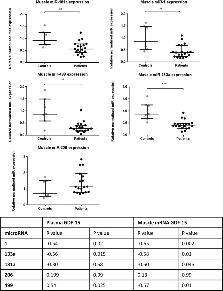
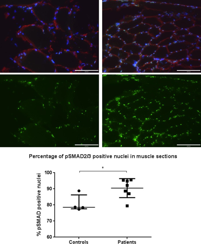
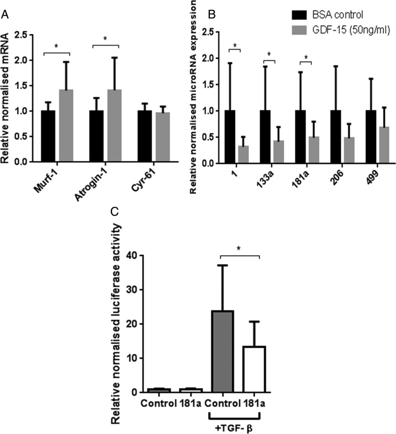
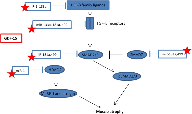
Measurements and main results: Compared with controls, GDF-15 protein was elevated in plasma (median 7239 vs 2454 pg/mL, p=0.001) and GDF-15 mRNA in the muscle (median twofold increase p=0.006) of patients with ICUAW. The expression of microRNAs involved in muscle homeostasis was significantly lower in the muscle of patients with ICUAW. GDF-15 treatment of C2C12 myotubes significantly elevated expression of muscle atrophy-related genes and down-regulated the expression of muscle microRNAs. miR-181a suppressed transforming growth factor-β (TGF-β) responses in C2C12 cells, suggesting increased sensitivity to TGF-β in ICUAW muscle. Consistent with this suggestion, nuclear phospho-small mothers against decapentaplegic (SMAD) 2/3 was increased in ICUAW muscle.

Conclusions: GDF-15 may increase sensitivity to TGF-β signalling by suppressing the expression of muscle microRNAs, thereby promoting muscle atrophy in ICUAW. This study identifies both GDF-15 and associated microRNA as potential therapeutic targets.

Keywords: Not Applicable; Respiratory Muscles.


Figures

Figure 1
Figure 1
Muscle biopsy specimens from the rectus femoris of ICUAW (n=7) and controls (n=4). H&E staining of muscle biopsies from control subjects (A) and patients (B) at 10× magnification. Immunostaining of the same control (C) and patient (D) for different muscle fibre types. Samples are representative of their respective groups. Blue, MHC 1; green, MHC 2a; red, laminin: type 2× fibres do not stain and can be seen as black fibres (10× magnification). (E) Mean fibre diameter of patients and controls. (F) Percentage distribution of all fibres measured (33–107 measured per subject). (G) Mean fibre type proportion of different MHCs 1, 2a, 2x and dual staining 1/2a fibres. (H) mRNA expression for different MHCs. Controls n=7, patients n=20; data presented as median and error bars represent IQR, *p<0.05, **p<0.01, ***p<0.001 Mann–Whitney. ICUAW, intensive care unit-acquired weakness; MHC, myosin heavy chain.
Figure 2
Figure 2
Growth and differentiation factor-15 (GDF-15) in patients with intensive care unit-acquired weakness patients (n=20) and controls (n=7) measured in plasma (A) and rectus femoris muscle biopsy mRNA expression (B). Dotted line in (A) represents 1200 pg/mL—the upper limit of normal plasma GDF-15. Data shown as median and IQR; **p<0.01, ***p<0.001—Mann–Whitney. Correlation of plasma GDF-15 with Sequential Organ Failure Assessment (SOFA) score at the time of sampling (C), r=Pearson's r value for correlation.
Figure 3
Figure 3
Rectus femoris muscle mRNA expression of different mRNA in patients with intensive care unit-acquired weakness (n=20) and controls (n=7) for atrogin and CYR61 (cytosine rich protein 61 (CYR61). Data presented as median and error bars represent IQR; **p<0.01, ***p<0.001 Mann–Whitney.
Figure 4
Figure 4
Rectus femoris muscle microRNA expression in patients with intensive care unit-acquired weakness (n=19) and controls (n=7). Table shows correlation of log (miR expression) with log (plasma growth and differentiation factor-15 (GDF-15)) and log (GDF-15 mRNA expression) Pearson’s r values and p values (Bonferroni corrected for multiple testing) are listed. Data presented as median and error bars represent IQR; **p<0.01, ***p<0.001, Mann–Whitney.
Figure 5
Figure 5
Phosphorylated small mothers against decapentaplegic 2/3 (p-SMAD 2/3) nuclear staining of muscle specimens for patients and controls. Images show control (left) and patient (right) 20× magnification muscle sections stained for p-SMAD2/3 localisation. Blue, 4′,6-diamidino-2-phenylindole nuclear; red, laminin; green, p-SMAD2/3. Lower images show p-SMAD2/3 fluorescence only of the same field of view, samples are representative of their respective groups. Graph shows percentage of pSMAD2/3-positive nuclei for controls (n=4) and patients (n=7). Data are presented as median and IQR, p=0.042, Mann–Whitney.
Figure 6
Figure 6
Effects of growth and differentiation factor-15 (GDF-15) or vehicle control on differentiated C2C12 myotubes. Day 8 differentiated C2C12 myotubes were treated with GDF-15 (50 ng/mL) or vehicle control (0.1% bovine serum albumin with 20 mM HCl) for 4 days, differential mRNA (A) and microRNA (B) expression was quantified (n=4 in triplicate). (C) Myoblasts were transfected with miR-181a or negative control, then CAGA-12 firefly and Renilla Luciferase plasmids. Following 6 h treatment with transforming growth factor-β (TGF-β) (2.5 ng/mL), relative luciferase activity was quantified (n=3 in triplicate). Data are normalised to their contemporary control. Data presented as mean and error bars represent SD; *p≤0.05, t test.
Figure 7
Figure 7
Schematic representation of the interaction between microRNAs and transforming growth factor-β (TGF-β) signalling. Stars represent those microRNA that maybe suppressed by growth and differentiation factor -15 (GDF-15) in intensive care unit-acquired weakness, resulting in a promotion of muscle atrophy. HDAC4, histone deacetylase 4; MuRF-1, Muscle Ring Finger-1; SMAD, small mothers against decapentaplegic.

References

    1. De Jonghe B, Sharshar T, Lefaucheur J, et al. . Paresis acquired in the intensive care unit: a prospective multicenter study. JAMA 2002;288:2859–67. - PubMed
    1. Batt J, dos Santos CC, Cameron JI, et al. . Intensive care unit-acquired weakness: clinical phenotypes and molecular mechanisms. Am J Respir Crit Care Med 2013;187:238–46. - PubMed
    1. Puthucheary ZA, Rawal J, McPhail M, et al. . Acute skeletal muscle wasting in critical illness. JAMA 2013;310:1591–600. - PubMed
    1. Bloch S, Polkey MI, Griffiths M, et al. . Molecular mechanisms of intensive care unit-acquired weakness. Eur Respir J 2012;39:1000–11. - PubMed
    1. Unsicker K, Spittau B, Krieglstein K. The multiple facets of the TGF-beta family cytokine growth/differentiation factor-15/macrophage inhibitory cytokine-1. Cytokine Growth Factor Rev 2013;24:373–84. - PubMed

Publication types

MeSH terms