. 2015 Apr;6(2):231-9.
doi: 10.1007/s13244-014-0372-y. Epub 2014 Dec 17.

Role of interventional radiology in the management of complications after pancreatic surgery: a pictorial review

Affiliations

Role of interventional radiology in the management of complications after pancreatic surgery: a pictorial review

Giovanni Mauri et al. Insights Imaging. 2015 Apr.

Abstract

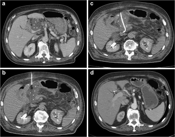
Pancreatic resections are surgical procedures associated with high incidence of complications, with relevant morbidity and mortality even at high volume centres. A multidisciplinary approach is essential in the management of these events and interventional radiology plays a crucial role in the treatment of patients developing post-surgical complications. This paper offers an overview on the interventional radiological procedures that can be performed to treat different type of complications after pancreatic resection. Procedures such as percutaneous drainage of fluid collections, percutaneous transhepatic biliary procedures, arterial embolisation, venous interventions and fistula embolisation are viable treatment options, with fewer complications compared with re-look surgery, shorter hospital stay and faster recovery. A selection of cases of complications following pancreatic surgery managed with interventional radiological procedure are presented and discussed. Teaching Points • Interventional radiology is crucial to treat complications after pancreatic surgery • Percutaneous drainage of collections can be performed under ultrasound or computed tomography guidance • Percutaneous biliary procedures can be used to treat biliary complications • Venous procedures can be performed effectively through transhepatic or transjugular access • Fistulas can be treated effectively by percutaneous embolisation.


Figures

Fig. 1
Fig. 1
Use of a percutaneous drainage to treat a post-pancreaticoduodenectomy fluid collection. a CT scan shows post-pancreaticoduodenectomy retroperitoneal fluid collection (asterisks). b The fluid collection (asterisks) is punctured with a small needle under CT guidance. c A percutaneous drainage (arrow) is inserted into the collection (asterisks) using the Seldinger technique. d At the end of the treatment complete resolution of the collection is obtained
Fig. 2
Fig. 2
Use of a percutaneous transhepatic biliary drainage to treat a post-surgical biliary leak. a Percutaneous colangiography demonstrates a biliary leak (arrow) and a biliary drainage (white arrows) is inserted via segment III left lobe approach; RBT right biliary tree, LBT left biliary tree, SB small bowel. b Final result with complete healing of the leak and absence of contrast leakage; RBT right biliary tree, LBT left biliary tree, SB bowel
Fig. 3
Fig. 3
Percutaneous treatment of a benign stricture of the common bile duct by a bioabsorbable biliary stent. a Percutaneous transhepatic cholangiography demonstrates remarkably dilated bile ducts (asterisks). The obstruction can be clearly spotted (white arrows). b Bilioplasty procedure. The balloon (black arrows) is fully inflated to dilate the stricture. Radiopaque contrast agent can be detected in the bowel (B); asterisks dilated bile ducts. c A bioabsorbable stent is positioned to keep the stricture open. The stent is radiolucent, but two platinum, radiopaque markers can be seen (black arrowheads); asterisks dilated bile ducts, B bowel. d The stent is fully expanded and radiopaque bile can be seen flowing through the patent common bile duct (white arrowheads); black arrows radiopaque stent markers; B bowel. e US follow-up at 3 months demonstrating good visibility of the stent (arrowheads) that was correctly expanded, and no dilation of the intrahepatic biliary ducts; L liver
Fig. 4
Fig. 4
Arterial embolisation of a post-surgical haemorrhage. a CT scan showing contrast extravasation (arrow) from the celiac trunk at the level of the bifurcation between hepatic and splenic artery; V inferior vena cava, A abdominal aorta. b Angiographic appearance of the active bleeding with contrast extravasation (arrows); asterisk celiac trunk, black arrowhead splenic artery, white arrowhead hepatic artery. c Angiography after embolisation with coils (arrows) demonstrating absence of contrast extravasation at the level of previous bleeding. d CT scan at 2 years, demonstrating absence of contrast extravasation at the level of previous bleeding and preserved patency of proper hepatic artery (white arrow), even in the presence of part of a coil in its lumen (arrowhead); black arrow clustered metallic coils in the focus of arterial lesion properly embolised
Fig. 5
Fig. 5
Treatment of a patient with postoperative mesenteric vein stenosis and thrombosis by percutaneous transhepatic stenting, thromboaspiration, and thrombolysis through a transjugular approach (the case has been partially previously reported [18]). a Maximum intensity projection reconstruction of abdominal MDCT shows surgical vascular graft (black arrow) at the level of the superior mesenteric vein and ascitic fluid (asterisks) b Percutaneous transhepatic angiography demonstrates stenosis at the level of proximal (black arrow) and distal (white arrow) anastomoses of the PTFE graft. c Angiography after stent placement (black arrow proximal stent, white arrow distal stent) demonstrating resolution of the stenosis. d MDCT MIP reconstruction and e axial image showing intra-stent thrombosis (white arrow) and ascitic fluid (asterisks). f Contrast injection in SMV (arrow) demonstrates complete vessel occlusion. g Angiography after aspiration thrombectomy and angioplasty shows residual thrombus (arrow). h Complete resolution and calibre restoration after direct thrombolysis with rt-PA
Fig. 6
Fig. 6
Successful percutaneous embolisation with cyanoacrilic glue of a postoperative fistula. a, b Contrast injection through a previously placed percutaneous drainage (white arrowhead) at the level of a fluid collection (asterisk) demonstrating a fistula (black arrow) with a communication (small black arrowhead) with the biliary system and bowel (B); large black arrowhead percutaneous transhepatic biliary drainage. c A guidewire (small white arrowhead) is advanced through the percutaneous drainage (large white arrowhead) into the fistula (black arrow); asterisk fluid collection, black arrowhead percutaneous transhepatic biliary drainage, B bowel. d A microcatheter (white arrowhead) is advanced over the wire at the level of the fistula (black arrow), and glue is injected (white arrow); asterisk fluid collection, black arrowhead percutaneous transhepatic biliary drainage, B bowel

References

    1. Buchler MW, Wagner M, Schmied BM, Uhl W, Friess H, Z’Graggen K (2003) Changes in morbidity after pancreatic resection: toward the end of completion pancreatectomy. Arch Surg 138(12):1310–1314 - PubMed
    1. Yeo CJ, Cameron JL, Sohn TA et al (1997) Six hundred fifty consecutive pancreaticoduodenectomies in the 1990s: pathology, complications, and outcomes. Ann Surg 226(3):248–257 - PMC - PubMed
    1. Sohn TA, Yeo CJ, Cameron JL et al (2000) Resected adenocarcinoma of the pancreas-616 patients: results, outcomes, and prognostic indicators. J Gastrointest Surg 4(6):567–579 - PubMed
    1. Gervais DA, Fernandez-del Castillo C, O’Neill MJ, Hahn PF, Mueller PR (2001) Complications after pancreatoduodenectomy: imaging and imaging-guided interventional procedures. Radiographics 21(3):673–690 - PubMed
    1. Zink SI, Soloff EV, White RR et al (2009) Pancreaticoduodenectomy: frequency and outcome of post-operative imaging-guided percutaneous drainage. Abdom Imaging 34(6):767–771 - PubMed