

Plasmodesmata of brown algae
- PMID: 25516500
- PMCID: PMC4375301
- DOI: 10.1007/s10265-014-0677-4
Plasmodesmata of brown algae
Abstract
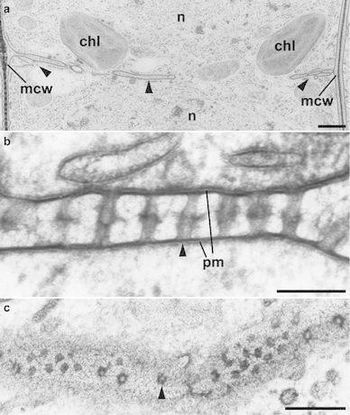
Plasmodesmata (PD) are intercellular connections in plants which play roles in various developmental processes. They are also found in brown algae, a group of eukaryotes possessing complex multicellularity, as well as green plants. Recently, we conducted an ultrastructural study of PD in several species of brown algae. PD in brown algae are commonly straight plasma membrane-lined channels with a diameter of 10-20 nm and they lack desmotubule in contrast to green plants. Moreover, branched PD could not be observed in brown algae. In the brown alga, Dictyota dichotoma, PD are produced during cytokinesis through the formation of their precursor structures (pre-plasmodesmata, PPD). Clustering of PD in a structure termed "pit field" was recognized in several species having a complex multicellular thallus structure but not in those having uniseriate filamentous or multiseriate one. The pit fields might control cell-to-cell communication and contribute to the establishment of the complex multicellular thallus. In this review, we discuss fundamental morphological aspects of brown algal PD and present questions that remain open.
Figures


Similar articles
-
Multicellularity and the Need for Communication-A Systematic Overview on (Algal) Plasmodesmata and Other Types of Symplasmic Cell Connections.Plants (Basel). 2023 Sep 21;12(18):3342. doi: 10.3390/plants12183342. Plants (Basel). 2023. PMID: 37765506 Free PMC article. Review.
-
Ultrastructural study of plasmodesmata in the brown alga Dictyota dichotoma (Dictyotales, Phaeophyceae).Planta. 2012 Oct;236(4):1013-26. doi: 10.1007/s00425-012-1656-4. Epub 2012 May 1. Planta. 2012. PMID: 22547029
-
Intercellular translocation of molecules via plasmodesmata in the multiseriate filamentous brown alga, Halopteris congesta (Sphacelariales, Phaeophyceae).J Phycol. 2017 Apr;53(2):333-341. doi: 10.1111/jpy.12498. Epub 2017 Jan 3. J Phycol. 2017. PMID: 27885652
-
Ultrastructural Observation of Cytokinesis and Plasmodesmata Formation in Brown Algae.Methods Mol Biol. 2022;2382:253-264. doi: 10.1007/978-1-0716-1744-1_16. Methods Mol Biol. 2022. PMID: 34705245
-
Polarization of brown algal zygotes.Semin Cell Dev Biol. 2023 Jan 30;134:90-102. doi: 10.1016/j.semcdb.2022.03.008. Epub 2022 Mar 19. Semin Cell Dev Biol. 2023. PMID: 35317961 Review.
Cited by
-
MUM, a maternal unknown message, inhibits early establishment of the medio-lateral axis in the embryo of the kelp Saccharina latissima.Development. 2024 Oct 15;151(20):dev202732. doi: 10.1242/dev.202732. Epub 2024 Sep 13. Development. 2024. PMID: 39190296 Free PMC article.
-
Multicellularity and the Need for Communication-A Systematic Overview on (Algal) Plasmodesmata and Other Types of Symplasmic Cell Connections.Plants (Basel). 2023 Sep 21;12(18):3342. doi: 10.3390/plants12183342. Plants (Basel). 2023. PMID: 37765506 Free PMC article. Review.
-
Three-dimensional growth: a developmental innovation that facilitated plant terrestrialization.J Plant Res. 2020 May;133(3):283-290. doi: 10.1007/s10265-020-01173-4. Epub 2020 Feb 24. J Plant Res. 2020. PMID: 32095969 Free PMC article. Review.
-
Plasmodesmata: function and diversity in plant intercellular communication. Preface.J Plant Res. 2015 Jan;128(1):3-5. doi: 10.1007/s10265-014-0697-0. J Plant Res. 2015. PMID: 25566753 No abstract available.
-
Distribution of natural ingredients suggests a complex network of metabolic transport between source and sink tissues in the brown alga Fucus vesiculosus.Planta. 2019 Feb;249(2):377-391. doi: 10.1007/s00425-018-3009-4. Epub 2018 Sep 12. Planta. 2019. PMID: 30209618
References
-
- Amat MA, Srivastava LM. Translocation of iodine in Laminaria saccharina (Phaeophyta) J Phycol. 1985;21:330–333. doi: 10.1111/j.0022-3646.1985.00330.x. - DOI
-
- Bauer R, Begerow D, Sampaio JP, Weiß M, Oberwinkler F. The simple-septate basidiomycetes: a synopsis. Mycol Prog. 2006;5:41–66. doi: 10.1007/s11557-006-0502-0. - DOI
-
- Bouget FY, Berger F, Brownlee C. Position dependent control of cell fate in the Fucus embryo: role of intercellular communication. Development. 1998;125:1999–2008. - PubMed
Publication types
MeSH terms
LinkOut - more resources
Full Text Sources
Other Literature Sources
