

Identification of Listeria monocytogenes determinants required for biofilm formation
- PMID: 25517120
- PMCID: PMC4269431
- DOI: 10.1371/journal.pone.0113696
Identification of Listeria monocytogenes determinants required for biofilm formation
Abstract
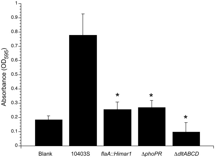
Listeria monocytogenes is a Gram-positive, food-borne pathogen of humans and animals. L. monocytogenes is considered to be a potential public health risk by the U.S. Food and Drug Administration (FDA), as this bacterium can easily contaminate ready-to-eat (RTE) foods and cause an invasive, life-threatening disease (listeriosis). Bacteria can adhere and grow on multiple surfaces and persist within biofilms in food processing plants, providing resistance to sanitizers and other antimicrobial agents. While whole genome sequencing has led to the identification of biofilm synthesis gene clusters in many bacterial species, bioinformatics has not identified the biofilm synthesis genes within the L. monocytogenes genome. To identify genes necessary for L. monocytogenes biofilm formation, we performed a transposon mutagenesis library screen using a recently constructed Himar1 mariner transposon. Approximately 10,000 transposon mutants within L. monocytogenes strain 10403S were screened for biofilm formation in 96-well polyvinyl chloride (PVC) microtiter plates with 70 Himar1 insertion mutants identified that produced significantly less biofilms. DNA sequencing of the transposon insertion sites within the isolated mutants revealed transposon insertions within 38 distinct genetic loci. The identification of mutants bearing insertions within several flagellar motility genes previously known to be required for the initial stages of biofilm formation validated the ability of the mutagenesis screen to identify L. monocytogenes biofilm-defective mutants. Two newly identified genetic loci, dltABCD and phoPR, were selected for deletion analysis and both ΔdltABCD and ΔphoPR bacterial strains displayed biofilm formation defects in the PVC microtiter plate assay, confirming these loci contribute to biofilm formation by L. monocytogenes.
Conflict of interest statement
Figures
References
-
- McCollum JT, Cronquist AB, Silk BJ, Jackson KA, O'Connor KA, et al. (2013) Multistate outbreak of listeriosis associated with cantaloupe. N Engl J Med 369:944–953. - PubMed
-
- (2013) Food microbiology: fundamentals and frontiers; Doyle MP, Beuchat LR, editors. Washington, D.C.: ASM Press. 1038 p.
-
- Costerton JW, Stewart PS, Greenberg EP (1999) Bacterial biofilms: a common cause of persistent infections. Science 21:1318–1322. - PubMed
Publication types
MeSH terms
Substances
Grants and funding
LinkOut - more resources
Full Text Sources
Other Literature Sources
