

Review
doi: 10.3390/biology3040892.
Hepatitis C virus life cycle and lipid metabolism
Affiliations
- PMID: 25517881
- PMCID: PMC4280516
- DOI: 10.3390/biology3040892
Item in Clipboard
Review
Hepatitis C virus life cycle and lipid metabolism
Biology (Basel).
.
Abstract
Hepatitis C Virus (HCV) infects over 150 million people worldwide. In most cases HCV infection becomes chronic, causing liver disease ranging from fibrosis to cirrhosis and hepatocellular carcinoma. HCV affects the cholesterol homeostasis and at the molecular level, every step of the virus life cycle is intimately connected to lipid metabolism. In this review, we present an update on the lipids and apolipoproteins that are involved in the HCV infectious cycle steps: entry, replication and assembly. Moreover, the result of the assembly process is a lipoviroparticle, which represents a peculiarity of hepatitis C virion. This review illustrates an example of an intricate virus-host interaction governed by lipid metabolism.
Figures

Genomic organization of HCV and protein synthesis. The HCV genome contains a single open reading frame flanked by 5ꞌ and 3ꞌ non-coding regions. The 5ꞌ NTR contains an internal ribosome entry site (IRES). After its synthesis, the HCV polyprotein is cleaved by host signal peptidase (red vertical arrows) and by viral encoded proteases (NS2 and NS3/4A) as indicated by corresponding arrows. An additional cleavage removing the carboxy-terminal region of the core protein is mediated by cellular signal peptide peptidase (green vertical arrow). The functions of the individual proteins are indicated in the text.
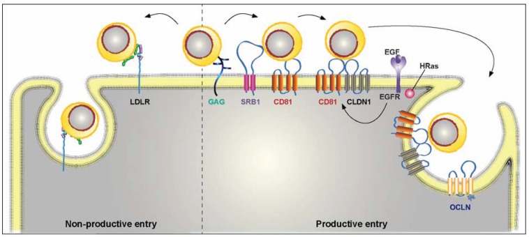
Cellular entry of HCV particles. HCV virion is associated with lipoproteins to form a complex particle that has been called lipoviroparticle (LVP). It initiates its life cycle by binding to glycosaminoglycans (GAGs). Then the virus can follow either a productive or a non-productive pathway. In the non-productive pathway, the lipoprotein component of the viral particle interacts with the LDL receptor (LDL-R) and the virion is rapidly internalized and potentially sent to a degradation pathway. The productive pathway is a complex multistep process involving a series of specific cellular entry factors, which include SRB1, CD81, tight-junction proteins, CLDN1 and OCLN, as well as other cellular factors not represented in this figure. After binding to several components of the host cell, HCV particle is internalized by clathrin-mediated endocytosis and fusion takes place in early endosomes.
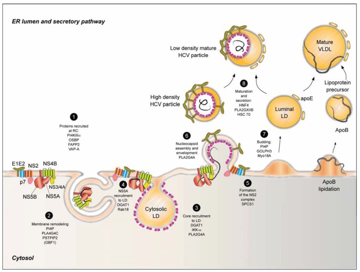
Model of viral replication and assembly of the infectious viral particle. Following viral protein processing, the mature proteins assemble in complexes. The NS2 complex consists of the envelope proteins E1 and E2, p7 viroporin and NS2 protease. The replication complex gather NS3 protease and its co-factor NS4A, NS4B, NS5A and NS5B, which are associated with modified membranes forming the “membranous web”. The cellular proteins PI4KIIIalpha, OSBP, FAPP2 and VAP-A are also recruited to the replication complexes, regulating membrane lipid exchange (1). NS4B protein induces the reorganization of intracellular membranes in special partially closed double membrane structures where replication occurs. The host proteins PLA2G4C and PSTPIP2 influence this membrane remodeling. Furthermore, GBF1 is involved in an early step of viral replication and it indirectly affects membrane remodeling (2). In the early stage of assembly core associates with LD following the recruitment of the replication complex through core-NS5A interaction. Several endogenous factors are involved in core and NS5A recruitment to LD and DGAT1 plays a central role in this relocalization (3 and 4). NS2 complexes (5) may interact with replication complexes accumulating in dotted structures in the proximity of LD. The presence of the three viral modules triggers the synchronous nucleocapsid assembly and envelopment potentiated by endogenous factors like PLA2G4A (6). Following the host-mediated budding (7), the immature viral particle fuses or attaches to a luminal lipid droplet through apoE-E1E2 interaction. The LVP takes a similar secretion and maturation route as VLDL, influenced by the cellular proteins HNF4, PLA2GXIIB and HSC70 (8).
References
Publication types
LinkOut - more resources
Full Text Sources
Other Literature Sources
