

Reliability of a 3 T MRI protocol for objective grading of supraspinatus tendonosis and partial thickness tears
- PMID: 25519001
- PMCID: PMC4278262
- DOI: 10.1186/s13018-014-0128-x
Reliability of a 3 T MRI protocol for objective grading of supraspinatus tendonosis and partial thickness tears
Abstract
Background: Partial thickness supraspinatus tears and tendonosis can be managed either nonoperatively or by various arthroscopic techniques. New biologic treatment approaches are currently being investigated. MRI is commonly used for objective imaging outcome evaluation but there is a lack of reliability studies. We propose a novel MRI classification of partial supraspinatus tears and tendonosis and evaluate its inter-observer and intra-observer reliability.
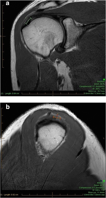
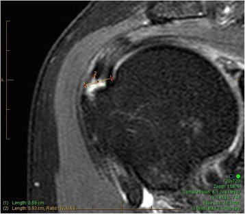
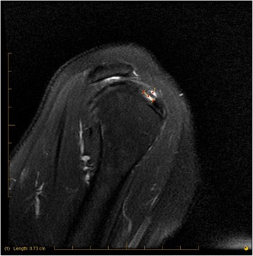
Methods: Digital MRI scans (3 Tesla) of 65 patients investigated for assessment of supraspinatus pathology or subacromial impingement were evaluated by three independent and experienced musculoskeletal (MSK) radiologists. Tendonosis (absent, focal, generalized), partial thickness (PT) tears (absent, 0%-25% PT, 25%-50% PT, 50%-100% PT, and full thickness tears), and anteroposterior extent of tears (less than 5 mm, 5-10 mm, greater than 10 mm) were scored by each radiologist on two separate occasions (t1, t2), 2 months apart. The inter-observer and intra-observer agreement and weighted kappa values for each parameter were calculated.
Results: The range of weighted intra-observer kappa (IAK) was 0.84-0.93 for evaluation of tendonosis; 0.84 (all raters) for depth of partial thickness, 0.74-0.84 for AP tear size, and 0.83-0.85 for the total score. The range of weighted inter-observer kappa (IEK) over two time points (t1, t2) was 0.55-0.74 for tendonosis, 0.69-0.84 for depth for partial thickness tears, 0.57-0.80 for AP tear size, and 0.63-0.80 for the total score.
Conclusion: A comprehensive MRI grading protocol is proposed and is reliable for the evaluation of supraspinatus tendonosis and partial thickness tears with good to excellent kappa values. This rotator cuff MRI protocol can be applied to evaluate morphological tendon outcomes after different treatment modalities.
Figures
References
-
- Khazzam M, Kuhn JE, Mulligan E, Abboud JA, Baumgarten KM, Brophy RH, Jones GL, Miller B, Smith M, Wright RW. Magnetic resonance imaging identification of rotator cuff retears after repair: interobserver and intraobserver agreement. Am J Sports Med. 2012;40(8):1722–1727. doi: 10.1177/0363546512449424. - DOI - PubMed
-
- Spencer EE, Jr, Dunn WR, Wright RW, Wolf BR, Spindler KP, McCarty E, Ma CB, Jones G, Safran M, Holloway GB, Kuhn JE. Interobserver agreement in the classification of rotator cuff tears using magnetic resonance imaging. Am J Sports Med. 2008;36(1):99–103. doi: 10.1177/0363546507307504. - DOI - PubMed
-
- Sher JS, Uribe JW, Posada A, Murphy BJ, Zlatkin MB. Abnormal findings on magnetic resonance images of asymptomatic shoulders. J Bone Joint Surg Am. 1995;77(1):10–15. - PubMed
Publication types
MeSH terms
LinkOut - more resources
Full Text Sources
Other Literature Sources
Medical
