. 2014 Dec 17:14:508.
doi: 10.1186/1472-6882-14-508.

Cytotoxicity effect of degraded and undegraded kappa and iota carrageenan in human intestine and liver cell lines

Affiliations

Cytotoxicity effect of degraded and undegraded kappa and iota carrageenan in human intestine and liver cell lines

Shahrul Hisham Zainal Ariffin et al. BMC Complement Altern Med. .

Abstract

Background: Carrageenan is a linear sulphated polysaccharide extracted from red seaweed of the Rhodophyceae family. It has broad spectrum of applications in biomedical and biopharmaceutical field. In this study, we determined the cytotoxicity of degraded and undegraded carrageenan in human intestine (Caco-2; cancer and FHs 74 Int; normal) and liver (HepG2; cancer and Fa2N-4; normal) cell lines.

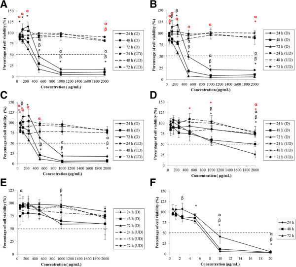
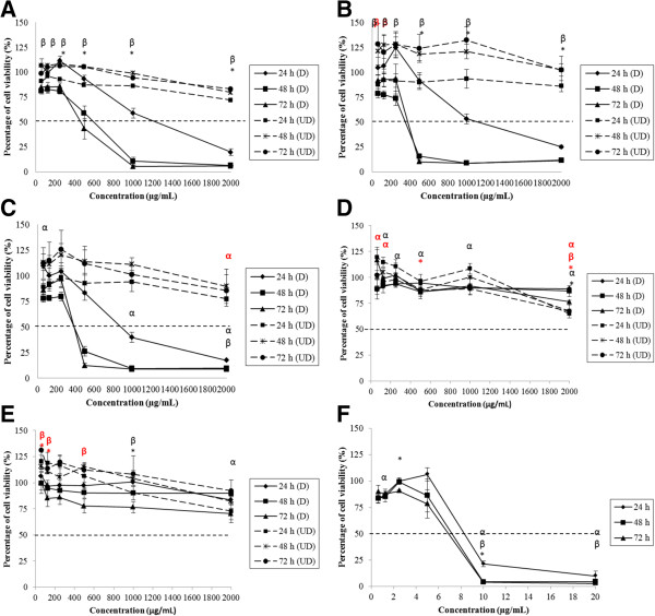
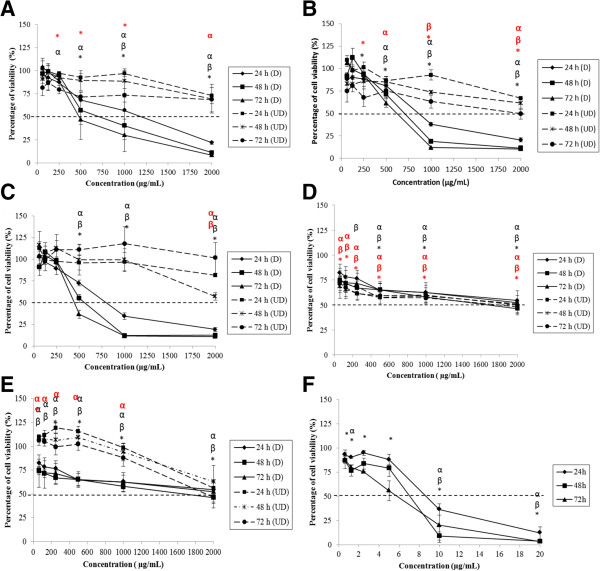
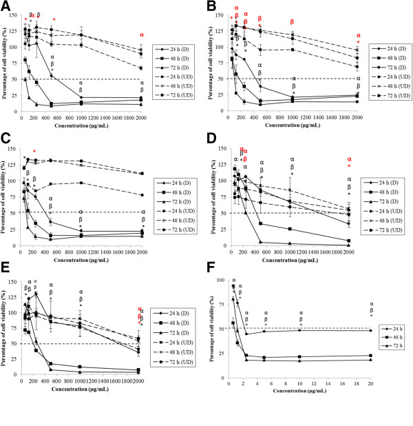
Methods: Food grade k-carrageenan (FGKC), dried sheet k-carrageenan (DKC), commercial grade k-carrageenan (CGKC), food grade i-carrageenan (FGIC) and commercial grade i-carrageenan (CGIC) were dissolved in hydrochloric acid and water to prepare degraded and undegraded carrageenan, respectively. Carrageenan at the concentration range of 62.5 - 2000.0 μg mL(-1) was used in the study. MTT assay was used to determine the cell viability while the mode of cell death was determined by May-Grunwald Giemsa (MGG) staining, acridine orange-ethidium bromide (AO/EtBr) staining, agarose gel electrophoresis and gene expression analysis.

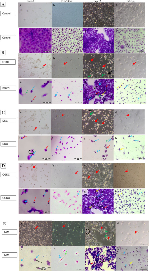
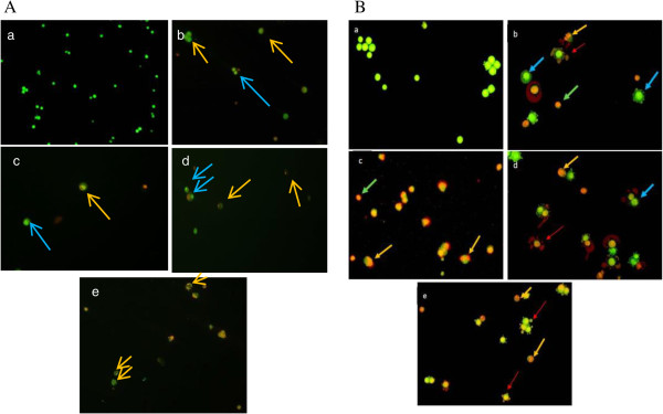
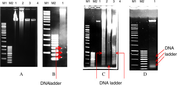
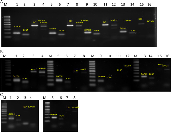
Results: Degraded FGKC, DKC and CGKC showed IC50 in 24, 48 and 72 hours treated Caco-2, FHs 74 Int, HepG2 and Fa2N-4 cell lines as tested by MTT assay. Degraded FGIC and CGIC only showed its toxicity in Fa2N-4 cells. The characteristics of apoptosis were demonstrated in degraded k-carrageenan treated Caco-2, FHs 74 Int, HepG2 and Fa2N-4 cells after MGG staining. When Caco-2 and HepG2 cells were undergone AO/EtBr staining, chromatin condensation and nuclear fragmentation were clearly seen under the microscope. However, DNA ladder was only found in HepG2 cells after gel electrophoresis analysis. Degraded k-carrageenan also inactivated PCNA, Ki-67 and survivin gene in HepG2. On the other hand, undegraded FGKC, DKC, CGKC, FGIC and CGIC treated cells showed no cytotoxic effect after analyzed by the same analyses as in degraded carrageenan.

Conclusion: Degraded k-carrageenan inhibited cell proliferation in Caco-2, FHs 74 Int, HepG2 and Fa2N-4 cell lines and the anti-proliferative effect was related to apoptosis together with inactivation of cell proliferating genes as determined by morphological observation and molecular analysis. However, no cytotoxic effect was found in undegraded carrageenan towards normal and cancer intestine and liver cell lines.


Figures

Figure 1
Figure 1
Concentration and time effects of degraded and undegraded carrageenan in Caco-2 cell line, as measured by the MTT assay. Caco-2 cells were treated with (A) FGKC, (B) DKC (C) CGKC (D) FGIC and (E) CGIC at concentrations of 62.5 – 2000.0 μg mL−1 and (F) tamoxifen at concentrations of 0.625- 20.000 μg mL−1 for 24 – 72 h. The percentages of cell viability were calculated by comparing the absorbance of treated and untreated cells. Degraded FGKC, DKC and CGKC showed IC50 in Caco-2 cells (black bold line) while IC50 values were not found in undegraded carrageenan treated cells (black dotted line). Each experiment had 3 replicates from 3 independent experiments (n = 3). The results are expressed as the mean ± SE. α: significant difference at 24 h; β: significant difference at 48 h and *: significant difference at 72 h for degraded carrageenan. Significant differences of undegraded carrageenan were in red symbols.
Figure 2
Figure 2
Concentration and time effects of degraded and undegraded carrageenan in FHs 74 Int cell line, as measured by the MTT assay. FHs 74 Int cells were treated with (A) FGKC, (B) DKC (C) CGKC (D) FGIC and (E) CGIC at concentrations of 62.5 – 2000.0 μg mL−1 and (F) tamoxifen at concentrations of 0.625- 20.000 μg mL−1 for 24 – 72 h. Degraded FGKC, DKC and CGKC showed IC50 in FHs 74 Int (black bold line). IC50 values were not found in degraded FGIC, CGIC and all undegraded carrageenan treated cells (black dotted line). Data are reported as means of three replicates from 3 independent experiments (n = 3). α: significant difference at 24 h; β: significant difference at 48 h and *: significant difference at 72 h for degraded carrageenan. Significant differences of undegraded carrageenan were in red symbols.
Figure 3
Figure 3
Concentration and time effects of degraded and undegraded carrageenan in HepG2 cell line, as measured by the MTT assay. HepG2 cells were subjected to the treatment of (A) FGKC, (B) DKC (C) CGKC (D) FGIC and (E) CGIC at concentrations of 62.5 – 2000.0 μg mL−1 and (F) tamoxifen at concentrations of 0.625- 20.000 μg mL−1 for 24 – 72 h. Degraded FGKC, DKC and CGKC also showed IC50 in HepG2 cells (black bold line). FGIC had IC50 > 1000 μg mL−1on the cells. Data are reported as means of three replicates from 3 independent experiments (n = 3). α: significant difference at 24 h; β: significant difference at 48 h and *: significant difference at 72 h for degraded carrageenan. Significant differences of undegraded carrageenan were in red symbols.
Figure 4
Figure 4
Concentration and time effects of degraded and undegraded carrageenan in Fa2N-4 cell line, as measured by the MTT assay. Fa2N4 cells were subjected to the treatment of (A) FGKC, (B) DKC (C) CGKC (D) FGIC and (E) CGIC at concentrations of 62.5 – 2000.0 μg mL−1 and (F) tamoxifen at concentrations of 0.625- 20.000 μg mL−1 for 24 – 72 h. All of the degraded carrageenan showing IC50 in Fa2N-4 cells (black bold line). Data are reported as means of three replicates from 3 independent experiments (n = 3). α: significant difference at 24 h; β: significant difference at 48 h and *: significant difference at 72 h for degraded carrageenan. Significant differences of undegraded carrageenan were in red symbols.
Figure 5
Figure 5
Morphology observations before and after MGG staining of Caco-2, FHs 74 Int, HepG2 and Fa2N-4 cells following exposure to degraded k -carrageenan. Cells were seeded at a cell density of 2 × 105 cells per ml and incubated for 24 h. A (a-h) Untreated cells. B (a-h) Cells treated with FGKC; 5C (a-h) Cells treated with DKC; 5D (a-h) Cells treated with CGKC and 5E (a-h) Cells treated with tamoxifen at the IC50 values for 72 h were observed for apoptotic morphological characteristics. Treated cells showed chromatin condensation (blue arrow), microspike formation (green arrow), nuclear fragmentation (yellow arrow) and apoptotic bodies (red arrow).
Figure 6
Figure 6
Caco-2 and HepG2 treated cells stained with acridine orange and ethidium bromide. Figure A shows Caco-2 cells while figure B shows HepG2 cells. (a) The untreated live cells showed normal green nuclei. (b) FGKC, (c) DKC, (d) CGKC and (e) tamoxifen-treated cells showed apoptosis, with bright green condensed nuclei and orange nuclei. Orange arrow: nucleus fragmentation. Blue arrow: chromatin condensation. Red arrow: apoptotic bodies. Light green arrow: necrotic cells.
Figure 7
Figure 7
Agarose gel electrophoresis of DNA extracted from untreated and treated HepG2 and Caco-2 cells. Panel A and B demonstrate results from Caco-2 while Panel C and D show results from HepG2 cells. Caco-2 treated cells did not show any DNA fragments ladder. The genomic DNA of untreated Caco-2 and HepG2 cells was intact, whereas DNA fragmentation with a ladder pattern of ~180 bp was found in all degraded k-carrageenan HepG2 cells. DNA of Caco-2 and HepG2 cells fragmented after induced by tamoxifen (Panel B and D). Lane M1 shows a 1 kb DNA ladder, and lane M2 shows a 100 kb DNA ladder. Panel A &C; lane 1: untreated cells, lane 2: degraded FGKC-treated cells, lane 3: degraded DKC-treated cells, lane 4: degraded CGKC-treated cells. Panel B &D; lane 1: Tamoxifen-treated cells.
Figure 8
Figure 8
Gene expression of proliferating cell nuclear antigen (PCNA), MKI67 and survivin analyzed by RT-PCR. (A) Gene expression of Caco-2 cells treated with degraded FGKC, DKC and CGKC. (B) Gene expression of HepG2 cells treated with degraded FGKC, DKC and CGKC. (C) Gene expression of Caco-2 and HepG2 cells treated with tamoxifen. GAPDH was used as the control gene. Degraded k-carrageenan was found inactivate the proliferating gene in HepG2 cells but not in Caco-2 cells. (A) &(B) Lane 1–4: untreated cells. Lane 5–8: FGKC. Lane 9–12: DKC and lane 13–16: CGKC. Lane 1, 5, 9 & 13: GAPDH. Lane 2,6,10% 14: PCNA. Lane 3,7,11 & 15: MKI67. Lane 4, 8, 12 & 16: Survivin. (C) Lane 1–4: Caco-2 cells. Lane 5–8: HepG2 cell.

References

    1. Tobacman JK. Review of harmful gastrointestinal effects of carrageenan in animal experiments. Environ Health Perspect. 2001;109:983. doi: 10.1289/ehp.01109983. - DOI - PMC - PubMed
    1. Meeting Joint FAO/WHO Expert Committee on Food Additives, Organization WH . Compendium of Food Additive Specifications: Addendum 9. 2001.
    1. Wang X, Xia Y, Liu L, Liu M, Gu N, Guang H, Zhang F. Comparison of MTT assay, flow cytometry, and RT‒PCR in the evaluation of cytotoxicity of five prosthodontic materials. J Biomed Mater Res B Appl Biomater. 2010;95:357–364. doi: 10.1002/jbm.b.31723. - DOI - PubMed
    1. Prajapati VD, Maheriya PM, Jani GK, Solanki HK. Carrageenan: a natural seaweed polysaccharide and its applications. Carbohydr Polym. 2014;105:97–112. doi: 10.1016/j.carbpol.2014.01.067. - DOI - PubMed
    1. Necas J, Bartosikova L. Carrageenan: a review. Vet Med. 2013;58:187–205.
Pre-publication history
    1. The pre-publication history for this paper can be accessed here:http://www.biomedcentral.com/1472-6882/14/508/prepub

Publication types

LinkOut - more resources