Comparative Study
. 2015 Feb 15;308(4):R305-20.
doi: 10.1152/ajpregu.00281.2014. Epub 2014 Dec 17.

Evolution of the oxygen sensitivity of cytochrome c oxidase subunit 4

Affiliations
Comparative Study

Evolution of the oxygen sensitivity of cytochrome c oxidase subunit 4

K M Kocha et al. Am J Physiol Regul Integr Comp Physiol. .

Abstract

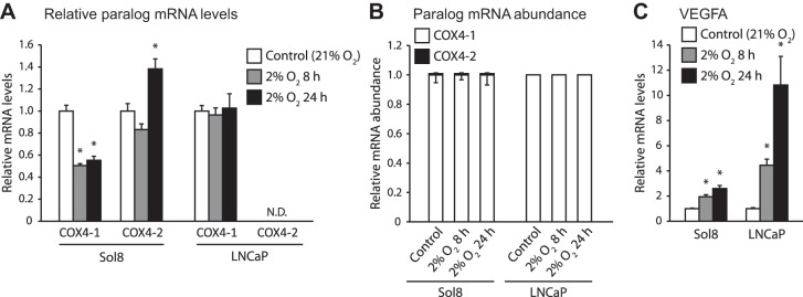
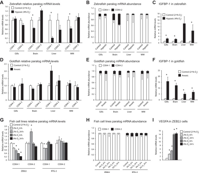
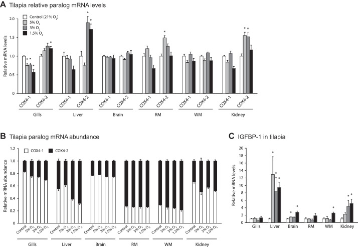
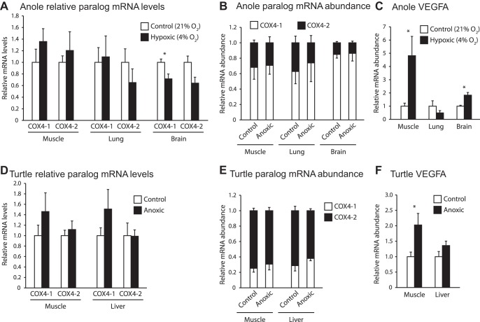
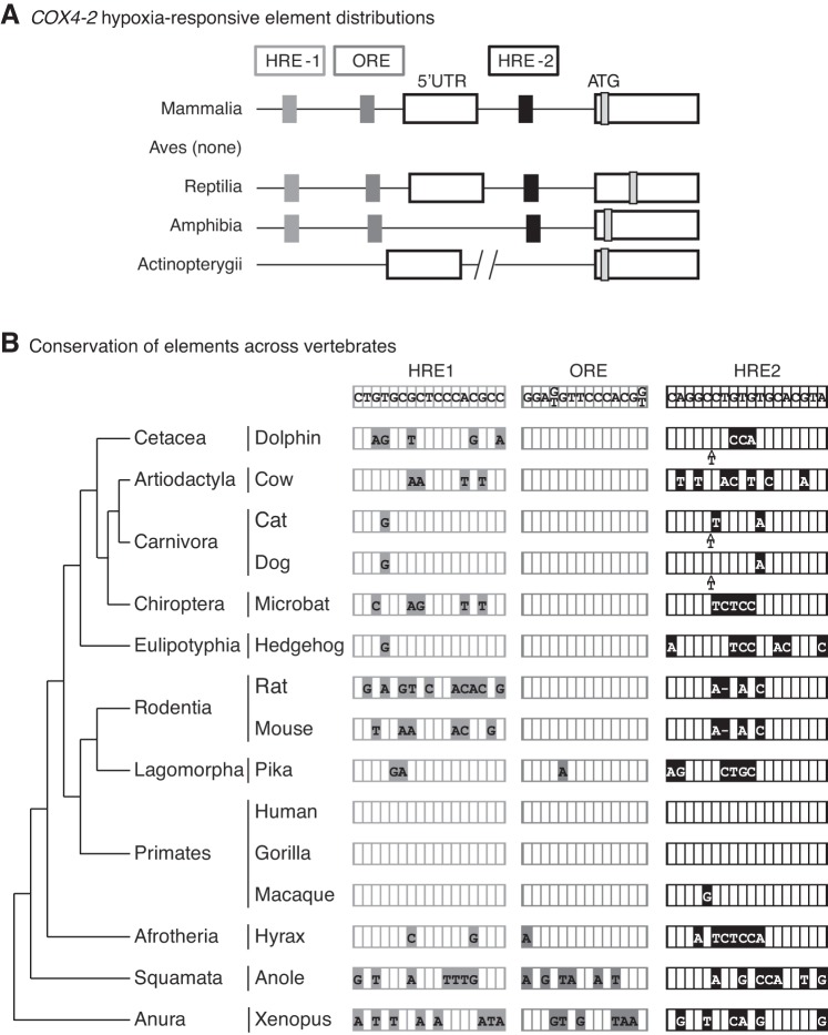
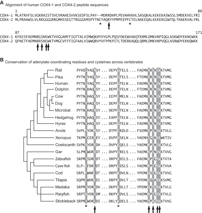
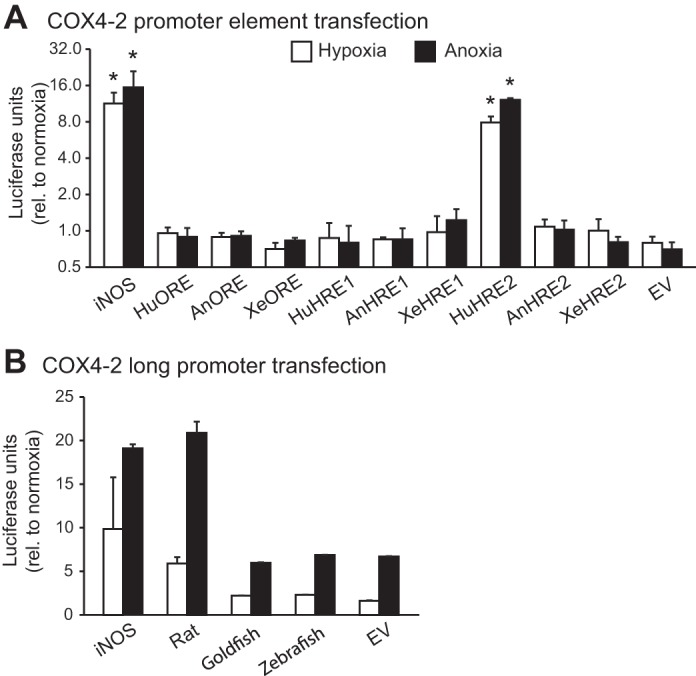
Vertebrates possess two paralogs of cytochrome c oxidase (COX) subunit 4: a ubiquitous COX4-1 and a hypoxia-linked COX4-2. Mammalian COX4-2 is thought to have a role in relation to fine-tuning metabolism in low oxygen levels, conferred through both structural differences in the subunit protein structure and regulatory differences in the gene. We sought to elucidate the pervasiveness of this feature across vertebrates. The ratio of COX4-2/4-1 mRNA is generally low in mammals, but this ratio was higher in fish and reptiles, particularly turtles. The COX4-2 gene appeared unresponsive to low oxygen in nonmammalian models (zebrafish, goldfish, tilapia, anoles, and turtles) and fish cell lines. Reporter genes constructed from the amphibian and reptile homologues of the mammalian oxygen-responsive elements and hypoxia-responsive elements did not respond to low oxygen. Unlike the rodent ortholog, the promoter of goldfish COX4-2 did not respond to hypoxia or anoxia. The protein sequences of the COX4-2 peptide showed that the disulfide bridge seen in human and rodent orthologs would be precluded in other mammalian lineages and lower vertebrates, all of which lack the requisite pair of cysteines. The coordinating ligands of the ATP-binding site are largely conserved across mammals and reptiles, but in Xenopus and fish, sequence variations may disrupt the ability of the protein to bind ATP at this site. Collectively, these results suggest that many of the genetic and structural features of COX4-2 that impart responsiveness and benefits in hypoxia may be restricted to the Euarchontoglires lineage that includes primates, lagomorphs, and rodents.

Keywords: COX4-2; HRE; ORE; oxygen sensitivity.


Figures

Fig. 1.
Fig. 1.
Transcriptional response to hypoxia in cytochrome c oxidase subunit 4 (COX4) paralogs in murine myoblast cells (Sol8) and human prostate cancer cells (LNCaP). Cells were exposed to 8 or 24 h of 2% O2; n = 6 per treatment. A: mRNA levels for COX4-1 and COX4-2, relative to control treatments. B: proportion of COX4 transcripts present as COX4-1 or COX4-2. C: relative mRNA levels for VEGFA, a known hypoxia-responsive gene. *Significant difference from the control treatment as determined by a Mann-Whitney U-test. Error bars depict SE.
Fig. 2.
Fig. 2.
Transcriptional response of COX4 paralogs to hypoxic treatments in whole zebrafish and goldfish, as well as ZEB2J and RTG-2 cell lines. A–C: transcriptional response in gills, brain, liver, and white muscle (WM) of zebrafish exposed to 8 h of 4% O2; n = 8 per treatment. D–F: transcriptional response in the corresponding tissues of goldfish exposed to 21.5 h of anoxia (undetectable levels of O2). G–I: transcriptional response of zebrafish epithelial cells (ZEB2J) and rainbow trout gonadal fibroblasts (RTG-2) exposed to 8 or 24 h of 2% O2 or anoxia; n = 6 per treatment. A, D, and G: mRNA levels for COX4-1 and COX4-2 relative to control treatments. B, E, and H: proportion of COX4 transcripts present as COX4-1 or COX4-2. C, F, and I: relative mRNA levels for insulin-like growth factor binding protein-1 (IGFBP-1; whole zebrafish and goldfish) or VEGFA (ZEB2J), which are known hypoxia-responsive genes. No response was seen in any hypoxia-responsive gene we measured in RTG-2 cells (data not shown). *Significant difference from the control treatment as determined by a Mann-Whitney U-test. Error bars depict SE.
Fig. 3.
Fig. 3.
Intertissue comparisons of the transcriptional response of COX4 paralogs to hypoxic treatments in tilapia. Transcriptional response in various tissues of tilapia exposed for 18 h to varying degrees of hypoxia; n = 6 per treatment, with exception of treatment group 1.5% where there was an n = 5 due to one mortality. A: mRNA levels for COX4-1 and COX4-2 relative to control treatments. B: proportion of COX4 transcripts present as COX4-1 or COX4-2. C: relative mRNA levels for IGFBP-1, a known hypoxia-responsive gene. *Significant difference from the control treatment as determined by a Mann-Whitney U-test. Error bars depict SE.
Fig. 4.
Fig. 4.
Time course of transcriptional response of COX4 paralogs to hypoxia in tilapia. Fish were exposed to hypoxia (5% O2) for 72 h, with single fish sampled throughout the time course. A: gill. B: liver. C: brain. D: red muscle. E: white muscle. F: kidney.
Fig. 5.
Fig. 5.
Transcriptional response of COX4 paralogs to hypoxic treatments in reptilian species. A–C: response in muscle, lung and brain tissues of green anole exposed to 4% O2 for 24 h. D–F: response in muscle and liver tissues of Western painted turtles exposed to 24 h or anoxia; n = 6 per treatment. A and D: mRNA levels for COX4-1 and COX4-2 relative to control treatments. B and E: proportion of COX4 transcripts present as COX4-1 or COX4-2. C and F: relative mRNA levels for VEGFA, a known hypoxia-responsive gene. *Significant difference from the control treatment as determined by a Mann-Whitney U-test. Error bars depict SE.
Fig. 6.
Fig. 6.
Comparison of putative hypoxia-responsive elements (HREs) in the COX4-2 gene across vertebrates. A and B: human sequences for the 2 HREs and the oxygen-responsive element (ORE) were used to search for orthologous elements in other vertebrate species representing several different lineages. A: location of the 3 elements in relation to the 5′-untranslated region (5′-UTR) and the ATG start site (ATG) of COX4-2 is shown for major classes of vertebrates. B determined elements are presented in relation to the vertebrate phylogeny. Differences from the consensus (human) sequence are indicated by filled boxes containing the alternative nucleotide or deletion (represented by a dash).
Fig. 7.
Fig. 7.
Conservation of features in vertebrate COX4-2 peptides. A: alignment of human COX4-1 and COX4-2 amino acid sequences. The COX4 amino acids suggested to be involved in the ATP/ADP binding site are denoted by arrows (Arg-20, Arg-73, Tyr-75, Glu-77, and Trp-78), and the COX4-2 cysteines that may disrupt the site are denoted by asterisks (Cys-16 and Cys-30). B: conservation of COX4-2 adenylate-coordinating amino acids and cysteines across vertebrates. Species names and accession numbers for each polypeptide sequence used can be found in Table 1.
Fig. 8.
Fig. 8.
Reporter gene activities for luciferase (pGL4.23) under control of the COX4-2 promoter and hypoxia responsive elements from candidate vertebrate species. A: PC3 cells were transfected with constructs containing 4 tandem repeats of HRE1 and 2 and the ORE from human (Hu), anole (An), and Xenopus (Xe), and were exposed to normoxia, 2% O2 (hypoxia), or anoxia for 24 h. B: HepG2 cells were transfected with constructs containing ∼2 kb of promoter sequence directly upstream of the COX4-2 5′-UTR from rat, goldfish, and zebrafish and were treated in the same way as the PC3 cells for A. Also included in both experiments were constructs containing four tandem repeats of a known hypoxia-responsive element from the human iNOS gene and the baseline activities for the empty pGL4.23 vector (EV). *Significant difference from the control treatment as determined by a Mann-Whitney U-test. Results were normalized to cotransfected Renilla reporter activity levels and are presented relative to normoxic activities (+SE).

References

    1. Aras S, Pak O, Sommer N, Finley R, Hüttemann M, Weissmann N, Grossman LI. Oxygen-dependent expression of cytochrome c oxidase subunit 4-2 gene expression is mediated by transcription factors RBPJ, CXXC5 and CHCHD2. Nucleic Acids Res 41: 2255–2266, 2013. - PMC - PubMed
    1. Arnold S. The power of life-cytochrome c oxidase takes center stage in metabolic control, cell signalling and survival. Mitochondrion 12: 46–56, 2012. - PubMed
    1. Arnold S, Kadenbach B. The intramitochondrial ATP/ADP-ratio controls cytochrome c oxidase activity allosterically. FEBS Lett 443: 105–108, 1999. - PubMed
    1. Bickler PE, Buck LT. Hypoxia tolerance in reptiles, amphibians, and fishes: life with variable oxygen availability. Annu Rev Physiol 69: 145–170, 2007. - PubMed
    1. Bremer K, Monk CT, Gurd BJ, Moyes CD. Transcriptional regulation of temperature-induced remodeling of muscle bioenergetics in goldfish. Am J Physiol Regul Integr Comp Physiol 303: R150–R158, 2012. - PubMed

Publication types

MeSH terms

LinkOut - more resources