. 2015 Feb 6;290(6):3666-79.
doi: 10.1074/jbc.M114.598797. Epub 2014 Dec 17.

The optimal corepressor function of nuclear receptor corepressor (NCoR) for peroxisome proliferator-activated receptor γ requires G protein pathway suppressor 2

Affiliations

The optimal corepressor function of nuclear receptor corepressor (NCoR) for peroxisome proliferator-activated receptor γ requires G protein pathway suppressor 2

Chun Guo et al. J Biol Chem. .

Abstract

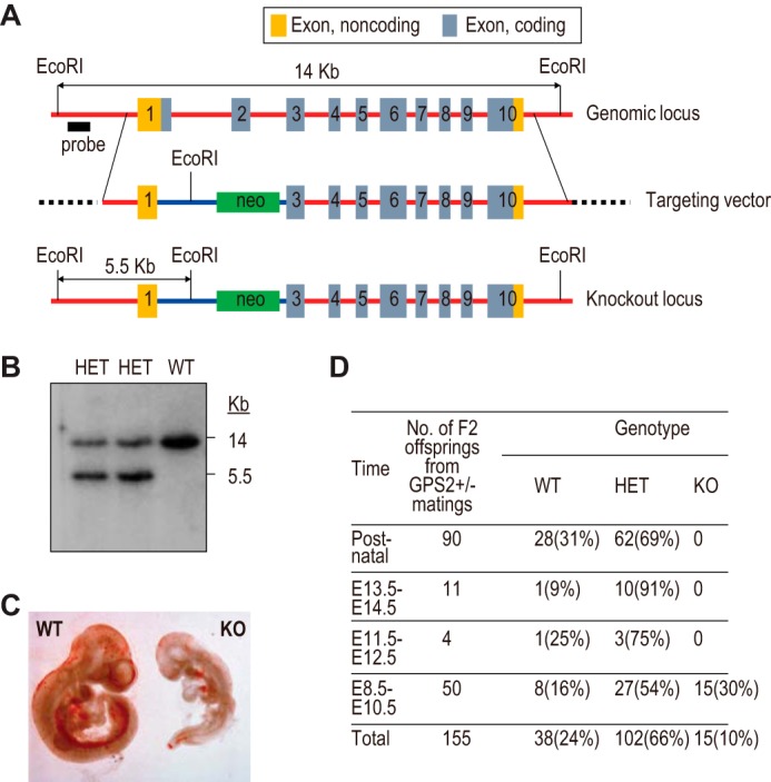
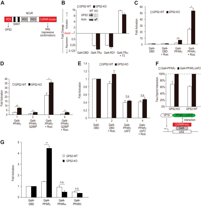
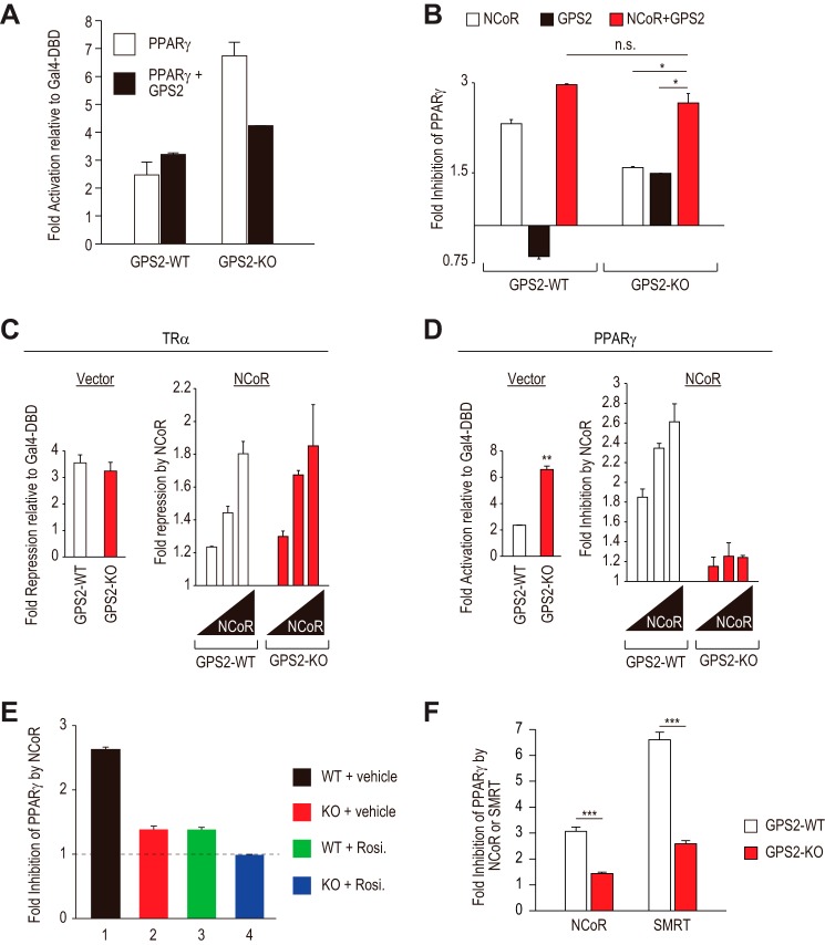
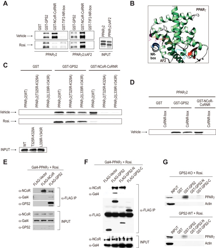
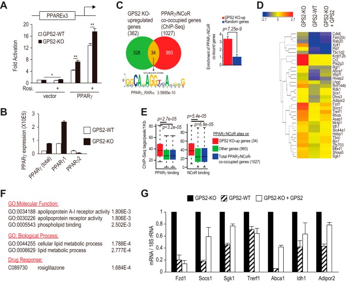
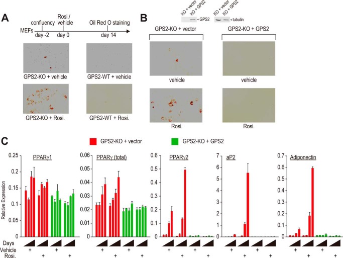
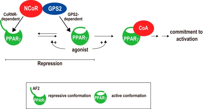
Repression of peroxisome proliferator-activated receptor γ (PPARγ)-dependent transcription by the nuclear receptor corepressor (NCoR) is important for homeostatic expression of PPARγ target genes in vivo. The current model states that NCoR-mediated repression requires its direct interaction with PPARγ in the repressive conformation. Previous studies, however, have shown that DNA-bound PPARγ is incompatible with a direct, high-affinity association with NCoR because of the inherent ability of PPARγ to adopt the active conformation. Here we show that NCoR acquires the ability to repress active PPARγ-mediated transcription via G protein pathway suppressor 2 (GPS2), a component of the NCoR corepressor complex. Unlike NCoR, GPS2 can recognize and bind the active state of PPARγ. In GPS2-deficient mouse embryonic fibroblast cells, loss of GPS2 markedly reduces the corepressor function of NCoR for PPARγ, leading to constitutive activation of PPARγ target genes and spontaneous adipogenesis of the cells. GPS2, however, is dispensable for repression mediated by unliganded thyroid hormone receptor α or a PPARγ mutant unable to adopt the active conformation. This study shows that GPS2, although dispensable for the intrinsic repression function of NCoR, can mediate a novel corepressor repression pathway that allows NCoR to directly repress active PPARγ-mediated transcription, which is important for the optimal corepressor function of NCoR for PPARγ. Interestingly, GPS2-dependent repression specifically targets PPARγ but not PPARα or PPARδ. Therefore, GPS2 may serve as a unique target to manipulate PPARγ signaling in diseases.

Keywords: Conformational Change; GPS2; NCoR; Nuclear Receptor; PPARγ; Peroxisome Proliferator-Activated Receptor (PPAR); Thyroid Hormone; Transcription Coactivator; Transcription Corepressor; Transcriptional Repression.


Figures

FIGURE 1.
FIGURE 1.
Embryonic lethality of whole-body GPS2 knockout mice. A, schematic of WT and KO GPS2 genomic loci and the design of the targeting vector to generate the whole-body GPS2 knockout mice. B, Southern blot analysis of WT and two independent heterozygous (HET) ES cell colonies. C, microscopic view of WT and KO embryos at E9.5. D, genotyping results of GPS2 WT, heterozygous, and KO mice at various embryonic developmental stages.
FIGURE 2.
FIGURE 2.
Loss of GPS2 derepresses PPARγ-dependent transcription. A, schematic of NCoR domains. RD1 interacts with GPS2. CoRNR boxes bind to NRs in the unliganded, repressive conformation. B, luciferase activities normalized to Gal4-DBD and assayed in GPS2-WT and GPS2-KO MEFs transfected with Gal4-RD1, Gal4-TRα (LBD), or the empty vector Gal4-DBD, along with a Gal4-UAS-driven luciferase reporter construct. A fold change of >1 denotes activation, whereas a fold change of <1 denotes repression. Inset, Western blot analysis of GPS2 expression in MEFs derived from GPS2-WT and GPS2-KO embryos. C, fold activation relative to Gal4-DBD assayed in GPS2-WT and GPS2-KO MEFs transfected with Gal4-PPARγ (LBD) or Gal4-DBD, along with a Gal4-UAS-driven luciferase reporter construct in the absence and presence of the PPARγ ligand rosiglitazone (Rosi.). *, p < 0.05; **, p < 0.01. D, similar to C, with inclusion of Gal4-PPARγQ286P (LBD). *, p < 0.05; **, p < 0.01. E, similar to C, except that Gal4-PPARγΔAF2 (LBD) was used in place of WT PPARγ. n.s., not significant. F, mammalian two-hybrid assays to measure the in vivo interactions between the NCoR CoRNR box region and PPARγ (LBD) or PPARγΔAF2 (LBD). Interactions derived from PPARγΔAF2 were set at 100%. G, GPS2 specifically inhibited PPARγ but not other PPAR isoforms. The experiments were similar to C, with inclusion of PPARα and PPARδ isoforms. **, p < 0.01. n.s., not significant.
FIGURE 3.
FIGURE 3.
GPS2 is important for PPARγ repression by NCoR. A, ectopic GPS2 restored PPARγ repression in GPS2-KO cells. Luciferase assays were performed in GPS2-WT and GPS-KO MEFs transfected with Gal4-PPARγ or empty vector, along with the Gal4-UAS reporter, in the absence or presence of GPS2. B, similar to A. GPS2-KO and WT MEFs were transfected with NCoR, GPS2, or both, as indicated. *, p < 0.05; n.s., not significant. C and D, NCoR showed defective corepressor function for PPARγ, but not TRα, in GPS2 KO MEFs. Luciferase assays were performed in GPS2 WT and GPS2 KO MEFs transfected with Gal4-PPARγ or Gal4-TRα, different doses of NCoR, or empty vector, along with the Gal4-UAS reporter. The left panels show basal levels of TRα-mediated repression (C) or PPARγ-mediated activation (D). Both were normalized to Gal4-DBD. The right panels show NCoR-elicited potentiation of TRα-dependent repression (C) or inhibition of PPARγ-dependent activation (D). **, p < 0.01. E, fold inhibition by NCoR of PPARγ transcriptional activity in GPS2 WT and GPS2 KO cells in the absence or presence of rosiglitazone (Rosi.). F, fold inhibition of PPARγ by NCoR or SMRT in GPS2 WT and GPS2 KO cells. 250 ng of NCoR and SMRT plasmids was used. The experiment was performed similar to D. ***, p < 0.001.
FIGURE 4.
FIGURE 4.
Differential recognition of PPARγ conformations by GPS2 and NCoR. A, GST pulldown assays to detect the in vitro interactions of PPARγ and PPARγΔAF2 with GPS2, NCoR, and TIF2 in the absence or presence of rosiglitazone (Rosi.). B, three-dimensional X-crystallographic structure of PPARγ in complex with rosiglitazone and an NR box peptide from SRC-1 (PDB code 2PRG) (6). It shows that the conserved Thr-325, Lys-329, Leu-339, and Val-343 residues located in the hydrophobic cavity directly contact the NR box peptide. C, mutation of the conserved hydrophobic cavity residues disrupted the PPARγ-NCoR interaction without significantly affecting the PPARγ-GPS2 interaction. D, CoRNR box peptide (100 μm) strongly inhibited the PPARγ-NCoR interaction but not the PPARγ-GPS2 interaction. E and F, coimmunoprecipitation assays performed in 293T cells transfected with FLAG-GPS2 or FLAG-NCoR, along with Gal4-PPARγ (LBD). The cells were cultured in the presence of rosiglitazone. Following anti-FLAG immunoprecipitation (IP), coimmunoprecipitated proteins were detected by Western blot analysis. G, GST pulldown assays were performed using cell lysates from GPS2 WT and GPS2 KO cells pretreated with rosiglitazone (1 μm) for 5 h. The results for WT and KO cells are from the same Western blot analysis but are presented separately.
FIGURE 5.
FIGURE 5.
Loss of GPS2 was sufficient to activate endogenous PPARγ target genes. A, luciferase assays measuring full-length PPARγ activity. GPS2 WT and GPS2 KO MEFs were transfected with a PPARγ responsive element (PPARE)-driven luciferase reporter along with full-length PPARγ or empty vector control in the absence and presence of rosiglitazone (Rosi.). *, p < 0.05; **, p < 0.01. B, RT-qPCR analysis of endogenous PPARγ1, PPARγ2, and total PPARγ in subconfluent GPS2 KO and GPS2 WT MEFs. C, left panel, overlap of genes up-regulated in GPS2 KO cells and genes physically bound by PPARγ and NCoR at the same site. RNA-Seq analysis was performed in GPS2 WT, GPS2 KO, and GPS2-re-expressed KO MEF cells. The mapped reads were analyzed for differential gene expression using DESeq and GeneSpring NGS software. Compared with GPS2-WT cells, 362 genes showed at least 2-fold higher expression in GPS2 KO but not in GPS2-re-expressed KO cells. The Venn diagram identified 34 overlapping genes between the 362 genes and genes containing overlapping binding sites of PPARγ and NCoR, which were determined by analyzing the ChIP-Seq data sets from mouse macrophages that express PPARγ (47, 48) using Homer (75). Left panel, bottom, de novo motif analysis (75) identified DR1 as the only enriched motif in the PPARγ binding sites on the 34 genes. Right panel, enrichment of PPARγ/NCoR target genes in GPS2 KO up-regulated genes, calculated as the ratio of the overlap with GPS2 KO up-regulated genes (i.e. 34 genes) versus random overlap with RefSeq genes (total of 37,593). 400 randomly generated RefSeq genes were used as a control. The p value was calculated on the basis of a binomial test. D, heatmap of the 34 genes in GPS2 WT, GPS2 KO, and GPS2-transduced KO cells. E, PPARγ and NCoR were present at higher levels on the 34 GPS2 KO up-regulated genes compared with genes not significantly up-regulated by the loss of GPS2. ChIP-Seq tags within a 1-kb region flanking PPARγ binding sites on genes co-occupied by PPARγ/NCoR were quantified using Homer. F, gene list enrichment analysis performed using the ToppGene server (76). GO, gene ontology. G, RT-qPCR analysis of gene expression in subconfluent GPS2 KO, GPS2 WT, and GPS2-re-expressed KO MEFs.
FIGURE 6.
FIGURE 6.
Loss of GPS2 converts MEFs into a preadipogenic state. A, Oil Red O staining of post-confluent GPS2 KO and GPS2 WT MEFs treated with vehicle or rosiglitazone (Rosi.). B, Oil Red O staining of post-confluent KO and GPS2-transduced KO MEFs treated with vehicle or rosiglitazone. Top panel, Western blot analysis of GPS2 expression in vector- and GPS2-transduced GPS2 KO cells. C, time course analyses of gene expression in post-confluent KO and GPS2-transduced KO cells by RT-qPCR at 0, 2, 4, and 6 days in the absence and presence of rosiglitazone.
FIGURE 7.
FIGURE 7.
Role of GPS2 in NCoR-mediated repression of PPARγ-dependent transcription. NCoR can directly regulate PPARγ not only in the repressive but also in the active conformation via CoRNR box-dependent and GPS2-dependent mechanisms, respectively. In the absence of agonists, a subset of PPARγ spontaneously adopts the active conformation that requires AF2, explaining why depleting AF2 insensitizes PPARγ to GPS2-dependent regulation. Agonists such as rosiglitazone increase the ability of PPARγ to assume the active state and to bind to CoAs. Unlike thought previously, repression occurs not only to the repressive state but also to the active state of PPARγ. See text for details.

References

    1. Mangelsdorf D. J., Thummel C., Beato M., Herrlich P., Schütz G., Umesono K., Blumberg B., Kastner P., Mark M., Chambon P., Evans R. M. (1995) The nuclear receptor superfamily: the second decade. Cell 83, 835–839 - PMC - PubMed
    1. Glass C. K., Rosenfeld M. G. (2000) The coregulator exchange in transcriptional functions of nuclear receptors. Genes Dev. 14, 121–141 - PubMed
    1. Bourguet W., Ruff M., Chambon P., Gronemeyer H., Moras D. (1995) Crystal structure of the ligand-binding domain of the human nuclear receptor RXR-α. Nature 375, 377–382 - PubMed
    1. Wagner R. L., Apriletti J. W., McGrath M. E., West B. L., Baxter J. D., Fletterick R. J. (1995) A structural role for hormone in the thyroid hormone receptor. Nature 378, 690–697 - PubMed
    1. Perissi V., Rosenfeld M. G. (2005) Controlling nuclear receptors: the circular logic of cofactor cycles. Nat. Rev. Mol. Cell Biol. 6, 542–554 - PubMed

Publication types

MeSH terms

Substances

Associated data