

The Sm protein methyltransferase PRMT5 is not required for primordial germ cell specification in mice
- PMID: 25519955
- PMCID: PMC4369312
- DOI: 10.15252/embj.201489319
The Sm protein methyltransferase PRMT5 is not required for primordial germ cell specification in mice
Abstract
PRMT5 is a type II protein arginine methyltransferase with roles in stem cell biology, reprograming, cancer and neurogenesis. During embryogenesis in the mouse, it was hypothesized that PRMT5 functions with the master germline determinant BLIMP1 to promote primordial germ cell (PGC) specification. Using a Blimp1-Cre germline conditional knockout, we discovered that Prmt5 has no major role in murine germline specification, or the first global epigenetic reprograming event involving depletion of cytosine methylation from DNA and histone H3 lysine 9 dimethylation from chromatin. Instead, we discovered that PRMT5 functions at the conclusion of PGC reprograming I to promote proliferation, survival and expression of the gonadal germline program as marked by MVH. We show that PRMT5 regulates gene expression by promoting methylation of the Sm spliceosomal proteins and significantly altering the spliced repertoire of RNAs in mammalian embryonic cells and primordial cells.
Keywords: PGC specification; PRMT5; conditional knockout; germline absence; splicing.
© 2014 The Authors.
Figures
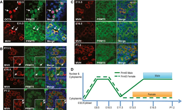
IF for PRMT5 (green) and OCT4+ PGCs (arrows) at E10.5, and MVH+ PGCs (arrows) at E11.5 (red).
PRMT5 (green) in male gonads at E13.5, E16.5 and P1-2. MVH (red) marks germ cells, with PRMT5 foci (white arrow heads) or MVH foci (red arrow heads).
PRMT5 (green) expression in female gonads at E13.5, E16.5 and P1-2. MVH (red) marks germ cells.
Summary of expression of PRMT5 in the mammalian germline.
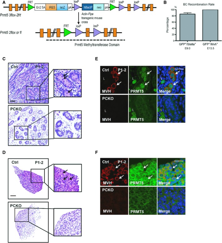
Schematic design of the targeting vector. Prmt53flox−2frt founders were mated with Actin-Flpe to excise the FRT flanked cassette to obtain Prmt52flox or Prmt5fl/+ mice. Prmt5fl/+ mice were intercrossed to obtain Prmt5fl/fl mice.
Recombination rate of Blimp1-Cre (BC). BC was crossed to YFP lox-stop-lox mice, and recombination rate was calculated based on the fraction of YFP+ cells in the STELLA+ (E9.0) or MVH+ (E13.5) fraction.
P1-2 male gonad (C) and P1-2 female gonad (D) in control (Ctrl) and PCKO embryos. Arrows indicate germ cells. Scale bar, 100 μm.
IF for PRMT5 (green) and MVH (red) in (E) P1-2 male and (F) P1-2 female gonads. L, Leydig cell. Arrows indicate germ cells. Scale bar, 20 μm.
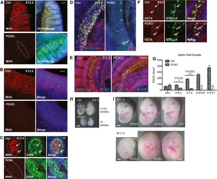
IF for MVH+ germline cells (red) in (A) male, and (B) female gonads at E13.5. In males, sertoli cells are also marked with SOX9 (green). Dashed circle indicates a testis cord. Scale bar, 50 μm.
Higher power image of (A) at E13.5. Dashed circle indicates a cross section through a testis cord. Arrows point to PGCs, which were identified by MVH+ staining. Scale bar, 20 μm.
IF of the indifferent gonads at E11.5 with PGCs identified by staining for STELLA (green) and MVH (red). Arrows indicate a small PGC cluster in the PCKO mutants. Scale bar, 100 μm. Note that PCKO mutant PGCs are STELLA-positive but not MVH-positive at this magnification.
IF at E9.5 with PGCs identified by co-staining for STELLA (green) and OCT4 (red). Scale bar, 100 μm.
Higher power image of (E) at E9.5. Arrows indicate OCT4 and STELLA double-positive PGCs. Scale bar, 20 μm.
Quantification of germ cell number between control and PCKO at different gestational stages.
E9.5 embryos from control and PCKO. Scale bar, 1 mm.
E11.5 embryos of control and PCKO. The majority of PCKO embryos are equivalent size to the control at E11.5. Scale bar, 1 mm.
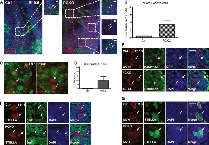
IF of E10.5 embryos showing OCT4+ (red) PGCs with cPARP (green). Arrows indicate apoptotic PGCs. Scale bar, 20 μm.
Quantification of apoptotic OCT4+ PGCs in control and PCKO embryos at E10.5. Data are shown as mean ± SEM. Standard error is across visual fields containing 10 PGCs. In total, about 4–5 fields were used for the quantification for each genotype.
IF of E9.5 embryos for Ki67 showing OCT4+ PGCs (arrows). Scale bar, 10 μm.
Quantification of Ki67 negative OCT4+ PGCs at E9.5. Data are shown as mean ± SEM.
IF at E10.5 for OCT4+ PGCs and H3K9me2. White arrows mark OCT4+ PGCs. Both Ctrl and PCKO PGCs show the absence of H3K9me2 (green) staining. Scale bar, 20 μm.
IF at E11.5 for STELLA+ PGCs and 5mC. White arrows mark STELLA+ PGCs. Both Ctrl and PCKO PGCs show the absence of global 5mC (green) staining. Note that STELLA+ PCKO PGCs are not MVH positive at this age. Scale bar, 20 μm.
IF at E11.5 for PGCs (arrows) with MVH (red) and STELLA (green). Scale bar, 20 μm.
Schematic model of ESC derivation. Recombination is induced with addition of 4-OHT in culture for 48 h.
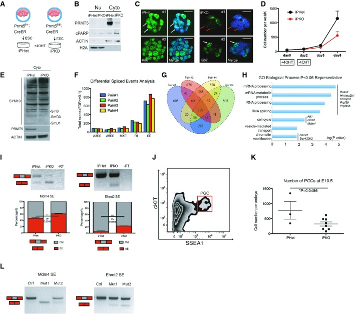
Western blot of iPHet and iPKO ESCs 5 days after treatment with 4-OHT. Nu, nuclear fraction. Cyto, cytoplasmic fraction. ACTIN and H2A are used for loading control of nuclear and cytoplasmic fraction, respectively.
IF for Ki67 (green) in iPHet and iPKO ESCs 5 days after treatment with 4-OHT. White arrows mark Ki67-positive ESCs. Red arrows mark Ki67-negative ESCs. Scale bar, 20 μm.
Growth curve of iPHet and iPKO ESCs at days 2, 3 and 5 after 4-OHT treatment. n = 2 replicates. The original seeding on day 0 is 50,000 (50K) cells per well of a 6-well plate.
Western blot for SYM10 antibody of iPHet and iPKO ESC cytoplasmic lysates 5 days after treatment with 4-OHT. Cyto, cytoplasmic fraction. ACTIN is used as a loading control. The absence of PRMT5 is shown in iPKO ESCs.
Pair-wise analysis of differential splicing events in each iPKO replicate relative to the paired iPHet control. A3SS, alternative 3′ splice site; A5SS, alternative 5′ splice site; MXE, mutual exclusive exons; RI, retained introns; SE, skipped exons.
Venn diagram of pair-wise analysis of MATS, showing overlapping differential splicing events among replicates.
Representative GO terms for common differentially spliced genes identified in three out of four replicate pairs.
Reverse transcript (RT) PCR validation of aberrant splicing events in Mdm4 and Ehmt2. SE, skipped exon. PCR is performed among at least three replicates for each locus. Electrophoresis image was quantified using ImageJ software. **P = 0.0046, ***P = 0.0006.
Representative flow plot of sorting cKIT+/SSEA1+ PGCs (red box) at E10.5 using FACS (shown is an embryo with genotype Prmt5fl/fl;creER with 4-OHT injection at E6.5).
Sorted PGC numbers among control iPHet (Prmt5fl/+;creER) and mutant iPKO (Prmt5fl/fl;creER). *P = 0.0486.
PCR validation of aberrant splicing events in Mdm4 and Ehmt2 using cDNA reverse-transcribed and amplified from FACS-sorted PGCs (individual embryos) using the NuGEN Ovation RNA Amp System V2. SE, skipped exon. PCR was performed among one control (Prmt5fl/+;creER) and two mutants (Prmt5fl/fl;creER).
Comment in
-
Prmt5: a guardian of the germline protects future generations.EMBO J. 2015 Mar 12;34(6):689-90. doi: 10.15252/embj.201591054. Epub 2015 Feb 16. EMBO J. 2015. PMID: 25687507 Free PMC article.
References
-
- Ancelin K, Lange UC, Hajkova P, Schneider R, Bannister AJ, Kouzarides T, Surani MA. Blimp1 associates with Prmt5 and directs histone arginine methylation in mouse germ cells. Nat Cell Biol. 2006;8:623–630. - PubMed
-
- Anne J, Ollo R, Ephrussi A, Mechler BM. Arginine methyltransferase Capsuleen is essential for methylation of spliceosomal Sm proteins and germ cell formation in Drosophila. Development. 2007;134:137–146. - PubMed
-
- Extavour CG, Akam M. Mechanisms of germ cell specification across the metazoans: epigenesis and preformation. Development. 2003;130:5869–5884. - PubMed
Publication types
MeSH terms
Substances
Grants and funding
LinkOut - more resources
Full Text Sources
Other Literature Sources
Molecular Biology Databases
