. 2015 Jan;43(1):618-28.
doi: 10.1093/nar/gku1270. Epub 2014 Dec 17.

Conformation transitions of eukaryotic polyribosomes during multi-round translation

Affiliations

Conformation transitions of eukaryotic polyribosomes during multi-round translation

Zhanna A Afonina et al. Nucleic Acids Res. 2015 Jan.

Abstract

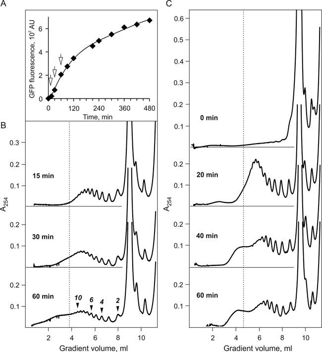
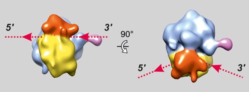
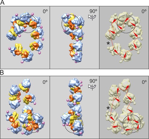
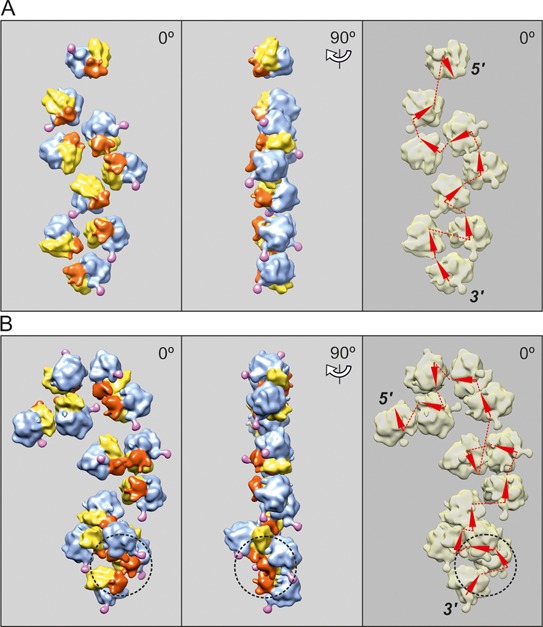
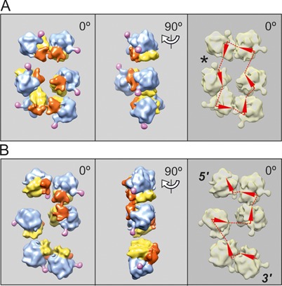
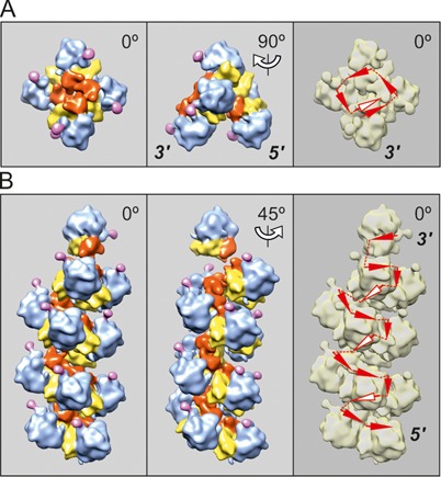
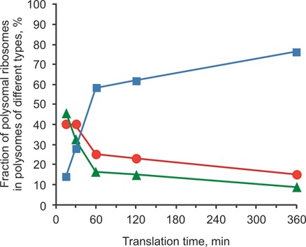
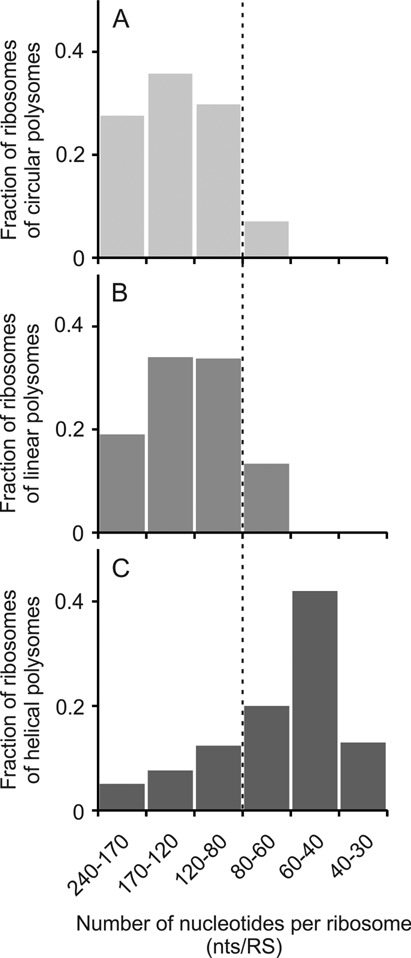
Using sedimentation and cryo electron tomography techniques, the conformations of eukaryotic polyribosomes formed in a long-term cell-free translation system were analyzed over all the active system lifetime (20-30 translation rounds during 6-8 h in wheat germ extract at 25°C). Three distinct types of the conformations were observed: (i) circular polyribosomes, varying from ring-shaped forms to circles collapsed into double rows, (ii) linear polyribosomes, tending to acquire planar zigzag-like forms and (iii) densely packed 3D helices. At the start, during the first two rounds of translation mostly the circular (ring-shaped and double-row) polyribosomes and the linear (free-shaped and zigzag-like) polyribosomes were formed ('juvenile phase'). The progressive loading of the polyribosomes with translating ribosomes induced the opening of the circular polyribosomes and the transformation of a major part of the linear polyribosomes into the dense 3D helices ('transitional phase'). After 2 h from the beginning (about 8-10 rounds of translation) this compact form of polyribosomes became predominant, whereas the circular and linear polyribosome fractions together contained less than half of polysomal ribosomes ('steady-state phase'). The latter proportions did not change for several hours. Functional tests showed a reduced translational activity in the fraction of the 3D helical polyribosomes.


Figures

Figure 1.
Figure 1.
Time course of protein synthesis (A) and polyribosomes formation (B and C) in the long-term cell-free translation system (CECF) programmed with 975-nt N-tagged GFP-encoding mRNA (A and B), or with 1650-nt luciferase-encoding mRNA (C) (see Materials and Methods, constructs 1 and 4, respectively). (A) Synthesis of GFP during 8 h of translation at 25°C. Accumulation of the protein during translation was recorded by measurement of the fluorescence intensity at 510 nm (with excitation at 395 nm). The arrows indicate the moments (15, 30 and 60 min) when the 25 μl samples of the translation mixture were taken for the sedimentation analysis. (B) Sucrose gradient sedimentation analysis of the polyribosomes formed in the cell-free translation system shown in (A): sedimentation profiles of polyribosomes formed after 15, 30 and 60 min translation (approximately corresponding to 2, 4 and 8 rounds of translation*, respectively). Zonal centrifugation was performed as follows: the 25 μl aliquots of the translation mixture were layered on the 15–45% sucrose gradient in 20 mM HEPES-KOH pH7.6, 5 mM magnesium acetate, 100 mM potassium acetate, 0.01 mg/ml cycloheximide buffer and centrifuged for 2 h at 37 000 rpm and 4°C in SW41 rotor. Sedimentation profiles were recorded by continuous measuring optical density at 254 nm in flow cell of Uvicord SII monitor during gradient fractionation. The vertical line on the sedimentation diagrams marks the position of dodecasome (12 translating ribosomes on 975 nt coding sequence, i.e. 80 nt per ribosome). (C) Time course of polyribosomes formation after 0, 20, 40 and 60 min of translation in CECF-system programmed with 1650 nt luciferase-encoding mRNA (construct 4). Sedimentation analysis of polyribosomes was performed in the same way as in (B) using 15–50% sucrose gradient. [*One round of translation is the time required for a ribosome to pass the full length of mRNA including initiation and termination.]
Figure 2.
Figure 2.
Structure of the eukaryotic (wheat) 80S ribosome obtained by averaging of the subtomograms resulted from cryo-ET analysis of polyribosomes within single tomogram. Here and in the Figures 3–6 the following colors are used: the head of the 40S ribosomal subunit is red, the body of the 40S subunit is yellow, the 60S subunit is blue and P1/P2 stalk is pink. The entry and exit sites of mRNA on the ribosome are indicated with dotted lines and arrowheads.
Figure 3.
Figure 3.
Cryo-ET reconstruction of circular polyribosomes. Nonasome (A) and undecasome (B) formed on uncapped non-adenylated GFP-encoding mRNA, 750-nt coding sequence (construct 2, see Materials and Methods). The ribosomes involved in 3D bulges (see the text) are inscribed into a dashed-line cycle. The undecasome (B) is an example of a double-row polyribosome with anti-parallel paths of the mRNA chain (collapsed circle). Here and in the following Figures 4–6, the left-hand panels present the vertical projections (elevation views) of the polyribosome reconstruction images. The middle panels show the side projections of the same polyribosome reconstruction. The right-hand panels present the deduced path of mRNA, the arrowheads being markers of the direction of mRNA path through the ribosomes. The asterisk indicates the presumed site of the junction of the 5′ and 3′ ends of mRNA, suggested by the gap and/or irregularity in orientation of neighboring ribosomes.
Figure 4.
Figure 4.
Cryo-ET reconstruction of topologically linear, zigzag-like polyribosomes. (A) Nonasome formed on uncapped non-polyadenylated GFP-encoding mRNA, 750-nt coding sequence (construct 2, see Materials and Methods). (B) Tridecasome formed on capped polyadenylated N-tagged GFP-encoding mRNA, 975-nt coding sequence (construct 1, see Materials and Methods). The nonasome (A) is an example of pseudo-double-row polyribosome with zigzag-like path of mRNA. The tridecasome in panel B contains a 3D bulge at its 3′-end (marked by a dashed-line cycle) with structural parameters similar to those of the turn of the dense 3D helical polyribosomes (see Discussion in the text).
Figure 5.
Figure 5.
Cryo-ET reconstruction of two topologically different hexasomes formed on capped polyadenylated N-tagged GFP-encoding mRNA, 975-nt coding sequence (construct 1, see Materials and Methods) after 15 min translation. (A) The hexasome with circular mRNA path, visible as short double-row polyribosome. (B) The hexasome with topologically linear, zigzag-like mRNA path, also visible as short polyribosome with two parallel rows of ribosomes (‘pseudo-double-row polysome’).
Figure 6.
Figure 6.
Cryo-ET reconstruction of 3D helical polyribosomes formed on capped polyadenylated N-tagged GFP-encoding mRNA, 975-nt coding sequence (construct 1, see Materials and Methods) after 30 min translation (A) and 120 min translation (B). (A) Pentasome folded into tetrad with one additional ribosome; the tetrad resembles the turn of the 4-fold helix as the typical conformation of the dense helical polyribosomes that appear at the late stages of the lifetime of translation (see below). (B) Tetradecasome folded into the dense 4-fold left-handed helix (see the text). The arrowheads indicating the mRNA path, which are pertinent to the ribosomes on the rear (invisible) side of helical polysomes, are marked by white color.
Figure 7.
Figure 7.
Dynamics of conformational changes of polyribosomes during multi-round translation in a long-term cell-free translation system programmed with the capped polyadenylated N-tagged GFP-encoding mRNA, 975 nt coding sequence (construct 1, see Materials and Methods): the distribution of polysomal ribosomes between different types of polyribosomes—circular (red circles), linear (green triangles) and 3D helical (blue squares)—depending on the time passed after translation start. The percentages of ribosomes in the polysomes of each type are plotted for each time point, the total number of polysomal ribosomes at this point being accepted as 100% (based on the data of Supplementary Table S1).
Figure 8.
Figure 8.
Dependence of proportions of three main types of polysomal conformations—circular (A), linear including zigzag-like (B) and 3D helical (C)—on the occupancy of the coding region of mRNA by translating ribosomes (number of nucleotides per ribosome, nts/RS ratio).

References

    1. Mathias A.P., Williamson R., Huxley H.E., Page S. Occurrence and function of polysomes in rabbit reticulocytes. J. Mol. Biol. 1964;9:154–167. - PubMed
    1. Dallner G., Siekevitz P., Palade G.E. Biogenesis of endoplasmic reticulum membranes. I. Structural and chemical differentiation in developing rat hepatocyte. J. Cell Biol. 1966;30:73–96. - PMC - PubMed
    1. Shelton E., Kuff E.L. Substructure and configuration of ribosomes isolated from mammalian cells. J. Mol. Biol. 1966;22:23–31.
    1. Christensen A.K., Kahn L.E., Bourne C.M. Circular polysomes predominate on the rough endoplasmic reticulum of somatotropes and mammotropes in the rat anterior pituitary. Am. J. Anat. 1987;178:1–10. - PubMed
    1. Yoshida T., Wakiyama M., Yazaki K., Miura K. Transmission electron and atomic force microscopic observation of polysomes on carbon-coated grids prepared by surface spreading. J. Electron Microsc. (Japan) 1997;46:503–506. - PubMed

Publication types