

Vesicle uncoating regulated by SH3-SH3 domain-mediated complex formation between endophilin and intersectin at synapses
- PMID: 25520322
- PMCID: PMC4328750
- DOI: 10.15252/embr.201439260
Vesicle uncoating regulated by SH3-SH3 domain-mediated complex formation between endophilin and intersectin at synapses
Abstract
Neurotransmission involves the exo-endocytic cycling of synaptic vesicle (SV) membranes. Endocytic membrane retrieval and clathrin-mediated SV reformation require curvature-sensing and membrane-bending BAR domain proteins such as endophilin A. While their ability to sense and stabilize curved membranes facilitates membrane recruitment of BAR domain proteins, the precise mechanisms by which they are targeted to specific sites of SV recycling has remained unclear. Here, we demonstrate that the multi-domain scaffold intersectin 1 directly associates with endophilin A to facilitate vesicle uncoating at synapses. Knockout mice deficient in intersectin 1 accumulate clathrin-coated vesicles at synapses, a phenotype akin to loss of endophilin function. Intersectin 1/endophilin A1 complex formation is mediated by direct binding of the SH3B domain of intersectin to a non-canonical site on the SH3 domain of endophilin A1. Consistent with this, intersectin-binding defective mutant endophilin A1 fails to rescue clathrin accumulation at neuronal synapses derived from endophilin A1-3 triple knockout (TKO) mice. Our data support a model in which intersectin aids endophilin A recruitment to sites of clathrin-mediated SV recycling, thereby facilitating vesicle uncoating.
Keywords: SH3 domains; endophilin; intersectin; neurotransmission; synaptic vesicle recycling.
© 2014 The Authors. Published under the terms of the CC BY NC ND 4.0 license.
Figures
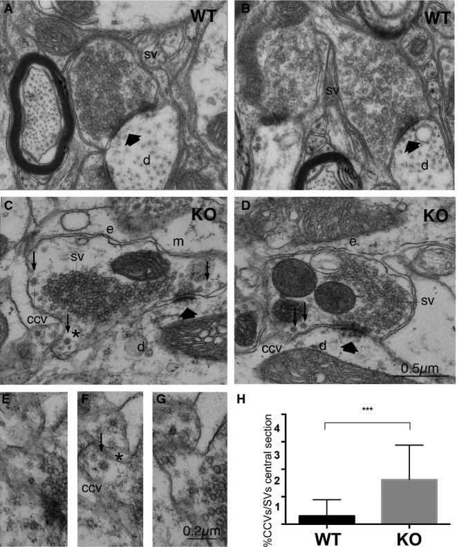
Electron micrographs of S-boutons establishing synapses on dendritic shafts in lamina IX of the mouse lumbar spinal cord of WT (A, B) or intersectin 1 KO mice (C, D). Scale bar: (A–D) 0.5 μm.
Serial section from area marked in (C) (asterisk); CCV, free clathrin-coated vesicle. Scale bar: (E–G) 0.2 μm.
Percentage of CCVs/total number of SVs (***P < 0.001; two-tailed unpaired t-test; n = 30, control and 32, intersectin 1 KO synapses from three mice of each genotype). Data are given as mean ± SD. SV, synaptic vesicles; d, dendritic shafts; e, endosome-like structures; m, mitochondrion; thick arrows indicate active zones; small arrows, CCVs.
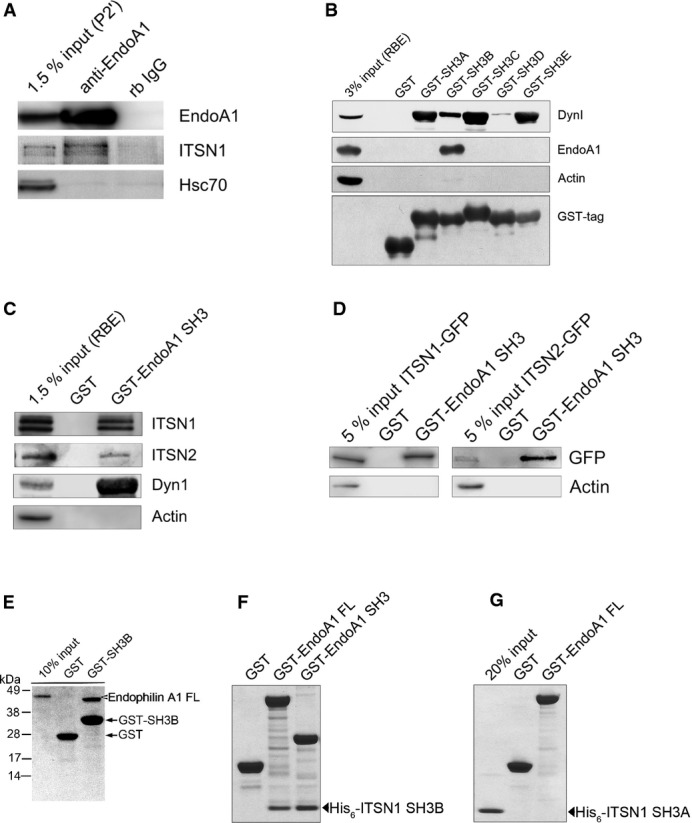
Endogenous endophilin A1 and intersectin 1 form a complex in situ. Immunoprecipitation from detergent-extracted rat brain synaptosomal fractions (P2′) using control rabbit non-immune IgG (rb IgG) or anti-endophilin A1 antibodies. Samples were analyzed by immunoblotting for endophilin A1 (EndoA1), intersectin 1 (ITSN1), and Hsc70.
GST-intersectin 1-SH3 associates with endophilin A1 present in detergent-lysed rat brain extract (RBE). Samples were analyzed by immunoblotting for endophilin A1 (EndoA1), dynamin 1 (DynI), actin, and GST.
Endophilin A1 associates with intersectin 2. (C) GST or GST-endophilin A1-SH3 fusion proteins were incubated with detergent-lysed rat brain extract (RBE). Samples were analyzed by immunoblotting for intersectin 1 (ITSN1), intersectin 2 (ITSN2; note: antibody specificity was verified by samples from intersectin 2 knockout mice), dynamin 1 (DynI), or actin. (D) Same as in (C) but using detergent extracts from HEK293 cells expressing intersectin 1-eGFP (ITSN1-GFP) or intersectin 2-eGFP (ITSN2-GFP). Samples were analyzed by immunoblotting for eGFP or actin.
Direct binding of endophilin A1-SH3 to intersectin 1-SH3B. Indicated GST-fusion proteins were incubated with full-length (FL) His6-endophilin A1 (E), His6-intersectin 1-SH3B (F), or His6-intersectin 1-SH3A (G). Samples were analyzed by SDS–PAGE and staining with Coomassie blue.
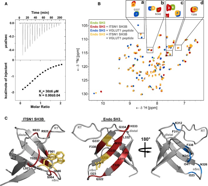
Isothermal titration calorimetry (ITC) profile of intersectin 1-SH3B titrated against endophilin A1-SH3. Top panel, heat changes upon ligand injection; the bottom panel, integrated power peaks fitted with a 1:1 model.
15N-HSQC spectrum of endophilin A1-SH3 (green) overlaid with spectra recorded after supplementation with intersectin 1-SH3B (red), a proline-rich peptide derived from VGLUT1 (blue) or both (yellow). Enlarged sections above illustrate typical signal changes caused by addition of SH3B (a) or peptide (b), both (c), or none (d).
Epitopes of the direct SH3-SH3 interaction mapped onto the structures of intersectin 1-SH3B (left) (PDB: 4IIM) and endophilin A1-SH3 (middle) (PDB:3IQL). Red, residues showing shift changes larger than the average shift distance (± SD) or disappearing due to line broadening upon SH3 domain binding. Blue, endophilin A1-SH3 residues that display significant shift perturbation after VGLUT1 peptide addition. Orange residues showed significant shift changes and were mutated in order to abrogate binding.
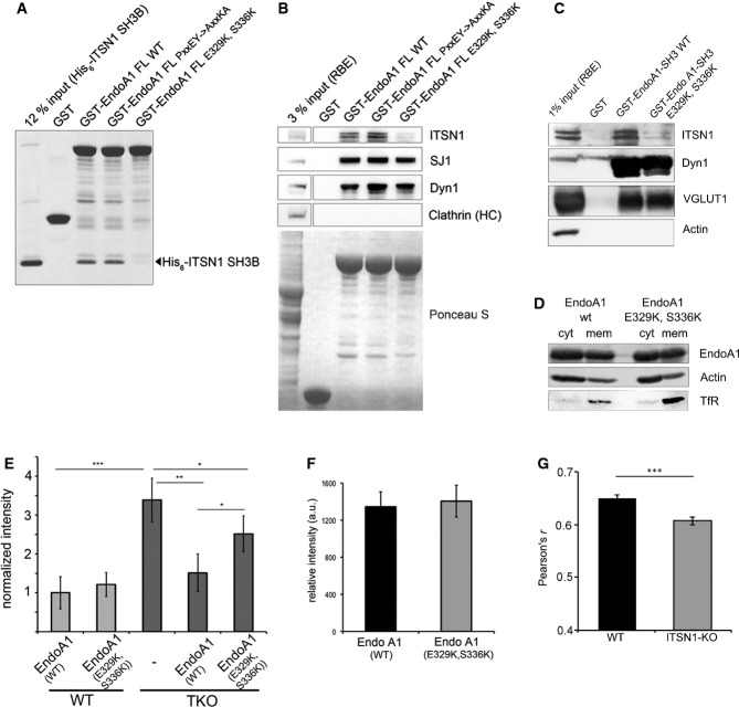
Endophilin A1 binding intersectin 1-SH3B binds via a non-canonical interface on its SH3 domain. GST-fused full-length (FL) endophilin A1 wild-type (WT), a proline-rich peptide binding defective mutant (PxxEY->AxxKA), or a mutant within the intersectin 1-SH3B binding interface (E329K, S336K) were incubated with purified recombinant intersectin 1-SH3B and analyzed by SDS–PAGE and staining with Coomassie blue.
Mutant endophilin A1 fails to bind to intersectin 1 while retaining association with proline-rich ligands. (B) GST or GST-fused full-length (FL) endophilin A1 wild-type (WT), a proline-rich peptide binding defective mutant (PxxEY->AxxKA), or a mutant within the intersectin 1-SH3B binding interface (E329K, S336K) were incubated with RBE. Samples were analyzed by immunoblotting for synaptojanin 1 (SJ1), dynamin 1 (Dyn1), or clathrin heavy chain (HC). (C) GST or GST-endophilin A1-SH3 wild-type (WT) or mutant (E329K, S336K) were incubated with RBE. Samples were analyzed by immunoblotting for intersectin 1 (ITSN1), vesicular glutamate transporter 1 (VGLUT1), dynamin 1 (Dyn1), or actin as a negative control.
Endophilin A1-mRFP WT and mutant (E329K, S336K) partition equally between membrane and soluble cytosolic fractions of HEK293 cells. Samples were immunoblotted for endophilin A1 (EndoA1), transferrin receptor (TfR), or actin.
Endophilin A1 binding to intersectin 1 regulates clathrin uncoating. Quantification of clathrin clustering in cortical neurons (DIV 14–22) from WT or endophilin A1-3 TKO mice re-expressing endophilin A1 WT or mutant (E329K, S336K). Clustering was fully rescued by endophilin A1-mRFP WT, but only to a minor degree by mutant endophilin A1 (E329K, S336K); y-axis, fold increase of fluorescence puncta in mutant synapses normalized to WT. *P < 0.05, **P < 0.01, ***P < 0.001, t-test.
Equal expression of endophilin 1 A1-mRFP wild-type (WT) or mutant (E329K, S336K) in primary cortical neurons (DIV 14–22; 68 random images from n = 4 independent experiments).
Intersectin 1 regulates endophilin A1 targeting to sites of clathrin-mediated endocytosis in neurons. Colocalization between endogenous endophilin A1 and AP-2 in primary hippocampal neurons (DIV 14) from wild-type (WT) or intersectin 1 knockout (KO) mice assessed by Pearson's correlation (four independent experiments; 111 for WT and 110 random images for KO). Data represent mean ± SEM. ***P < 0.0001, two-tailed unpaired t-test.
References
-
- Dittman J, Ryan TA. Molecular circuitry of endocytosis at nerve terminals. Annu Rev Cell Dev Biol. 2009;25:133–160. - PubMed
-
- Haucke V, Neher E, Sigrist SJ. Protein scaffolds in the coupling of synaptic exocytosis and endocytosis. Nat Rev Neurosci. 2011;12:127–138. - PubMed
-
- Daumke O, Roux A, Haucke V. BAR domain scaffolds in dynamin-mediated membrane fission. Cell. 2014;156:882–892. - PubMed
Publication types
MeSH terms
Substances
LinkOut - more resources
Full Text Sources
Other Literature Sources
Molecular Biology Databases
