Review
. 2014 Dec 3:8:392.
doi: 10.3389/fnins.2014.00392. eCollection 2014.

"You Shall Not Pass"-tight junctions of the blood brain barrier

Affiliations
Review

"You Shall Not Pass"-tight junctions of the blood brain barrier

Hans-Christian Bauer et al. Front Neurosci. .

Abstract

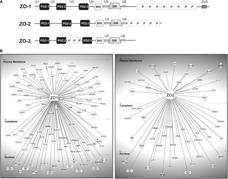
The structure and function of the barrier layers restricting the free diffusion of substances between the central nervous system (brain and spinal cord) and the systemic circulation is of great medical interest as various pathological conditions often lead to their impairment. Excessive leakage of blood-borne molecules into the parenchyma and the concomitant fluctuations in the microenvironment following a transient breakdown of the blood-brain barrier (BBB) during ischemic/hypoxic conditions or because of an autoimmune disease are detrimental to the physiological functioning of nervous tissue. On the other hand, the treatment of neurological disorders is often hampered as only minimal amounts of therapeutic agents are able to penetrate a fully functional BBB or blood cerebrospinal fluid barrier. An in-depth understanding of the molecular machinery governing the establishment and maintenance of these barriers is necessary to develop rational strategies allowing a controlled delivery of appropriate drugs to the CNS. At the basis of such tissue barriers are intimate cell-cell contacts (zonulae occludentes, tight junctions) which are present in all polarized epithelia and endothelia. By creating a paracellular diffusion constraint TJs enable the vectorial transport across cell monolayers. More recent findings indicate that functional barriers are already established during development, protecting the fetal brain. As an understanding of the biogenesis of TJs might reveal the underlying mechanisms of barrier formation during ontogenic development numerous in vitro systems have been developed to study the assembly and disassembly of TJs. In addition, monitoring the stage-specific expression of TJ-associated proteins during development has brought much insight into the "developmental tightening" of tissue barriers. Over the last two decades a detailed molecular map of transmembrane and cytoplasmic TJ-proteins has been identified. These proteins not only form a cell-cell adhesion structure, but integrate various signaling pathways, thereby directly or indirectly impacting upon processes such as cell-cell adhesion, cytoskeletal rearrangement, and transcriptional control. This review will provide a brief overview on the establishment of the BBB during embryonic development in mammals and a detailed description of the ultrastructure, biogenesis, and molecular composition of epithelial and endothelial TJs will be given.

Keywords: MAGUK proteins; PDZ scaffolds; blood-brain barrier; brain capillary endothelial cells; cell polarity; tight junctions; vascular permeability.


Figures

Figure 1
Figure 1
Establishment of the intraneural vascular plexus and a functional BBB during mouse embryogenesis. (A) Semithin section of embryonic mouse neocortex at E10 showing the intraneural and parts of the surrounding perineural domain (B) neuroepithelial cells surround a primitive capillary (white arrow) containing a red blood cell (asterisk). Murine E13 (C) and E17 (D) embryos microperfused with 0.5% Trypan blue solution. After fixation specimens were cut medially and photographed. Arrowhead indicates and enlarged area depicts stained choroid plexus (Bauer et al., 1993, 1995).
Figure 2
Figure 2
Vascular lumen formation by cord hollowing. (A) Intercellular adhesion of two or more ECs is accomplished by homophilic interaction of VE-cadherin and Pecam-1. (B) Following establishment of Par3-mediated apicobasal polarity via ß-1 integrin matrix interaction and the adaptor protein RASIP1, CD34-sialomucins (CD34 and podocalyxin; PODXL) are recruited to cell-cell contacts delineating the future apical border. VE-cadherin is required for specifically targeting CD34-sialomucins to contact sites, and the delivery depends on PTEN-mediated transformation of PIP3 to PIP2. Rasip1 localizes at inter-endothelial junctions and acts downstream of Rap1 (Ras-related protein1), mediating EC-ECM adhesion and stabilizing junctional integrity by actin bundling. In cooperation with Radil (ras-association and dilute domain-containing protein), Rasip1 further inhibits Rho-mediated stress fiber formation, thereby increasing endothelial barrier function. Further, CCM1 mediates the stabilization of β-catenin-containing endothelial cell-cell junctions downstream of the Rap1 GTPase. (C) Due to the negatively charged CD34-sialomucins, electrostatic repulsion leads to separation of the contacting plasma membranes thereby creating a free (non-contacting) surface and a small lumen between ECs. Concomitantly, cadherin-based AJs are redistributed to lateral borders, indicating the establishment of a lateral junctional complex. Following phosphorylation by protein kinase C, pMoesin, the major ERM protein in endothelial cells, is recruited to the future apical membrane and links CD34-sialomucins to F-actin, supporting cytoskeletal rearrangements in ECs during lumen expansion. Further, non-muscle myosin assembles at the apical plasma membrane and interacts with F-actin in response to VEGF signaling to support lumen expansion and cell shape changes. (D) Further maturation of the endothelial TJs is then mediated by claudin-5, occludin, members of the junctional adhesion molecule (JAM) family, or by EC-selective adhesion molecule (ESAM).
Figure 3
Figure 3
VE-cadherin and CCM1 act in concert to establish endothelial polarization and lumen formation. Rap1 regulates the junctional localization of CCM1 and mediates CCM1 activity in endothelial polarization and lumen formation. CCM1 stabilizes VE-cadherin at AJs, by associating with ß-catenin and inhibiting dissociation of ß-catenin from AJs. Thereby, nuclear accumulation of ß-catenin and ß-catenin transcriptional signaling is impaired. VE-cadherin promotes the apical localization and activation of the Par3 polarity complex. While VE-cadherin is needed for phosphorylation of PKCξ, CCM1/Rap1 is responsible for the junctional recruitment of the polarity complex. The accumulation of podocalyxin (PODXL) at the presumptive apical membrane is controlled by VE-cadherin and ß1 integrin. PODXL in turn recruits moesin and filamentous actin (F-actin) to the apical domain initiating endothelial lumen formation.
Figure 4
Figure 4
Transmembrane proteins of the TJ. (A) Cerebral endothelial cells constitute the cellular building block of the BBB and control the para- and transcellular routes via elaborate influx and efflux transporters and cell-cell adhesion structures. Astroglial endfeet, neurons, and microglia which are present at the neurovascular unit have been omitted for clarity. (B) Schematic membrane-spanning models of the transmembrane components present at TJs.
Figure 5
Figure 5
TJ-MAGUKS are membrane-associated adaptor proteins assembling the tight junction plaque. (A) ZO-1, ZO-2, and ZO-3 are multi-domain proteins composed of three PDZ domains, one SH3 and a catalytically inactive GUK domain. Next to these MAGUK-related domains, ZO proteins also contain various ZO-specific unique (U) and proline-rich (P) regions. ZU5 is a region found in ZO-1 and Unc5-like netrin receptors. (B) ZO-1 and ZO-2 are at the basis of large protein assemblies containing structural and signaling molecules. Shown are proteins that have been demonstrated to interact with ZO-1 or ZO-2 in epithelial and endothelial cells. Gene symbols according to the H.G.N.C. nomenclature are indicated.

References

    1. Abbott N. J. (2002). Astrocyte-endothelial interactions and blood-brain barrier permeability. J. Anat. 200, 629–638. 10.1046/j.1469-7580.2002.00064.x - DOI - PMC - PubMed
    1. Abbott N. J. (2013). Blood-brain barrier structure and function and the challenges for CNS drug delivery. J. Inherit. Metab. Dis. 36, 437–449. 10.1007/s10545-013-9608-0 - DOI - PubMed
    1. Abbott N. J., Ronnback L., Hansson E. (2006). Astrocyte-endothelial interactions at the blood-brain barrier. Nat. Rev. Neurosci. 7, 41–53. 10.1038/nrn1824 - DOI - PubMed
    1. Adachi M., Inoko A., Hata M., Furuse K., Umeda K., Itoh M., et al. (2006). Normal establishment of epithelial tight junctions in mice and cultured cells lacking expression of ZO-3, a tight-junction MAGUK protein. Mol. Cell. Biol. 26, 9003–9015. 10.1128/MCB.01811-05 - DOI - PMC - PubMed
    1. Adamson P., Etienne S., Couraud P. O., Calder V., Greenwood J. (1999). Lymphocyte migration through brain endothelial cell monolayers involves signaling through endothelial ICAM-1 via a rho-dependent pathway. J. Immunol. 162, 2964–2973. - PubMed