. 2014 Dec 1:6:328.
doi: 10.3389/fnagi.2014.00328. eCollection 2014.

The extraocular muscle stem cell niche is resistant to ageing and disease

Affiliations

The extraocular muscle stem cell niche is resistant to ageing and disease

Luigi Formicola et al. Front Aging Neurosci. .

Abstract

Specific muscles are spared in many degenerative myopathies. Most notably, the extraocular muscles (EOMs) do not show clinical signs of late stage myopathies including the accumulation of fibrosis and fat. It has been proposed that an altered stem cell niche underlies the resistance of EOMs in these pathologies, however, to date, no reports have provided a detailed characterization of the EOM stem cell niche. PW1/Peg3 is expressed in progenitor cells in all adult tissues including satellite cells and a subset of interstitial non-satellite cell progenitors in muscle. These PW1-positive interstitial cells (PICs) include a fibroadipogenic progenitor population (FAP) that give rise to fat and fibrosis in late stage myopathies. PICs/FAPs are mobilized following injury and FAPs exert a promyogenic role upon myoblasts in vitro but require the presence of a minimal population of satellite cells in vivo. We and others recently described that FAPs express promyogenic factors while satellite cells express antimyogenic factors suggesting that PICs/FAPs act as support niche cells in skeletal muscle through paracrine interactions. We analyzed the EOM stem cell niche in young adult and aged wild-type mice and found that the balance between PICs and satellite cells within the EOM stem cell niche is maintained throughout life. Moreover, in the adult mdx mouse model for Duchenne muscular dystrophy (DMD), the EOM stem cell niche is unperturbed compared to normal mice, in contrast to Tibialis Anterior (TA) muscle, which displays signs of ongoing degeneration/regeneration. Regenerating mdx TA shows increased levels of both PICs and satellite cells, comparable to normal unaffected EOMs. We propose that the increase in PICs that we observe in normal EOMs contributes to preserving the integrity of the myofibers and satellite cells. Our data suggest that molecular cues regulating muscle regeneration are intrinsic properties of EOMs.

Keywords: Duchenne muscular dystrophy; PICs; extraocular muscles; muscle stem cell niche; satellite cells.


Figures
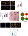
Figure 1
Figure 1
Wild-type EOM stem cell niche is intrinsically different from limb muscles. (A) Cross-sections of 7-week old EOMs (upper panels) and TA (lower panels) from C57Bl6 mice stained for PW1 (green) and the satellite cell marker Pax7 (red). Laminin staining (orange) shows the basal lamina. Nuclei were counterstained by DAPI (blue). Arrows indicate PICs, arrowheads indicate satellite cells. Scale bar, 50 µm. (B) Number of satellite cells and PICs per 100 fibers in 7-week old EOMs and TA cross-sections as stained in (A) revealed a bigger amount of PICs but not satellite cells in EOMs compared to TA. (C) Fold change of FST, IGF-1 and myostatin expression levels from qPCR analysis on total RNA extracts from EOMs and TA from 7 week-old wild-type mice revealed an strong increase in FST expression in EOMs. For TA muscles, three different animals (n = 3) were considered separately; for EOMs, six different animals (n = 6) were pooled into one sample. Each sample was analyzed in duplicate. Error bar indicates s.e.m. calculated for number of samples. (D) Xgal staining on cross-sections of 7-week old (upper panels) and 18 month-old (lower panels) EOMs and TA from PW1nLacZ mice. Nuclei and myofibers were counterstained with Nuclear Fast Red™ Solution. Scale bar, 40 µm. (E) Ratio between PICs (green) and satellite cells (red) per 100 fibers in 7-week old and 18-month old TA and EOMs cross-sections demonstrated that EOM but not TA stem cell niche is retained throughout life. (F) Cross-sections of 7-week old EOMs stained as in (A). We observe Pax7pos cells totally or partially surrounded by the basal lamina and often co-expressing PW1. Arrows indicate double-labeled PW1posPax7pos interstitial cells. (G) Number of Pax7pos interstitial cells (PW1pos and PW1neg subsets) per 100 fibers in 7 week-old and 18 month-old EOMs and TA cross-sections stained as in panel A. For all graphs, values represent the mean number of cells ± s.e.m. For (B,E), PICs were determined as interstitial PW1posPax7neg cells, satellite cells were determined as Pax7pos cells underneath the basal lamina. Statistical significance was calculated from at least three animals of each condition. *p < 0.05 **p < 0.01 ***p < 0.001.
Figure 2
Figure 2
Extraocular muscles from mdx and wild-type mice exhibit a similar muscle stem cell niche. (A) Cross-sections of 7-week old TA (upper panels) and EOMs (lower panels) from mdx and age-matched wild-type mice stained with hematoxylin and eosin showed large regenerating areas in TA but not EOMs from mdx mice. (B,C) Number of centrally nucleated fibers (B) and interstitial nuclei (C) per 100 fibers indicated the presence of an ongoing regeneration process in mdx TA but not EOMs. (D,E) Number of satellite cells per 100 sublaminal nuclei (D) and PICs per 100 interstitial nuclei (E) revealed an activation of both progenitor cell types in TA but not EOMs from mdx mice, as compared to their wild-type counterparts. Positive interstitial cells were determined as interstitial PW1posPax7neg cells, satellite cells were determined as Pax7pos cells underneath the basal lamina. For all graphs, values represent the mean number ± s.e.m. Statistical significance was calculated from at least three animals per each condition. *p < 0.05 **p < 0.01 ***p < 0.001.

References

    1. Abe S., Soejima M., Iwanuma O., Saka H., Matsunaga S., Sakiyama K., et al. . (2009). Expression of myostatin and follistatin in Mdx mice, an animal model for muscular dystrophy. Zoolog. Sci. 26, 315–320. 10.2108/zsj.26.315 - DOI - PubMed
    1. Abu-Baker A., Rouleau G. A. (2007). Oculopharyngeal muscular dystrophy: recent advances in the understanding of the molecular pathogenic mechanisms and treatment strategies. Biochim. Biophys. Acta 1772, 173–185. 10.1016/j.bbadis.2006.10.003 - DOI - PubMed
    1. Amthor H., Nicholas G., McKinnell I., Kemp C. F., Sharma M., Kambadur R., et al. . (2004). Follistatin complexes Myostatin and antagonises Myostatin-mediated inhibition of myogenesis. Dev. Biol. 270, 19–30. 10.1016/s0012-1606(04)00118-6 - DOI - PubMed
    1. Besson V., Smeriglio P., Wegener A., Relaix F., Nait Oumesmar B., Sassoon D. A., et al. . (2011). PW1 gene/paternally expressed gene 3 (PW1/Peg3) identifies multiple adult stem and progenitor cell populations. Proc. Natl. Acad. Sci. U S A 108, 11470–11475. 10.1073/pnas.1103873108 - DOI - PMC - PubMed
    1. Bismuth K., Relaix F. (2010). Genetic regulation of skeletal muscle development. Exp. Cell Res. 316, 3081–3086. 10.1016/j.yexcr.2010.08.018 - DOI - PubMed

LinkOut - more resources