. 2014 Nov 19;4(6):683-97.
eCollection 2014.

SPRY1 promotes the degradation of uPAR and inhibits uPAR-mediated cell adhesion and proliferation

Affiliations

SPRY1 promotes the degradation of uPAR and inhibits uPAR-mediated cell adhesion and proliferation

Xiufeng Liu et al. Am J Cancer Res. .

Abstract

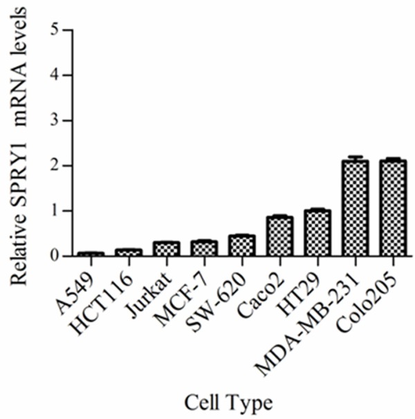
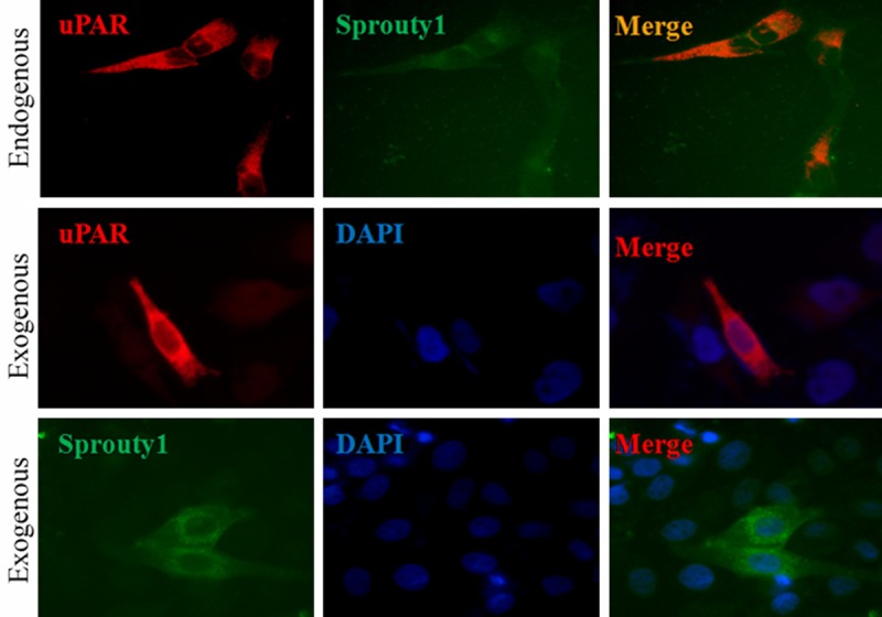
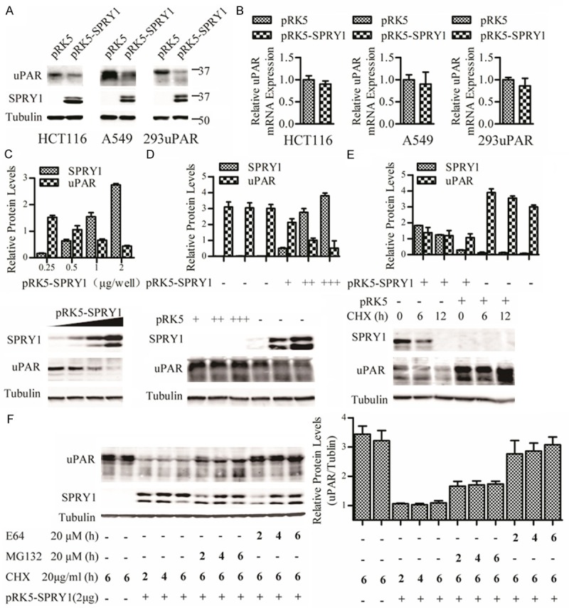
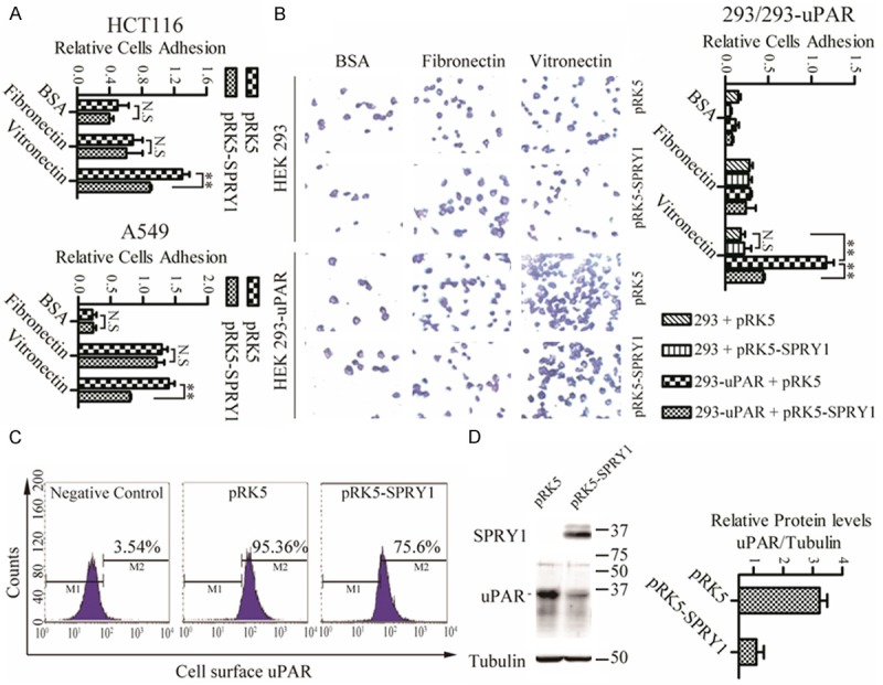
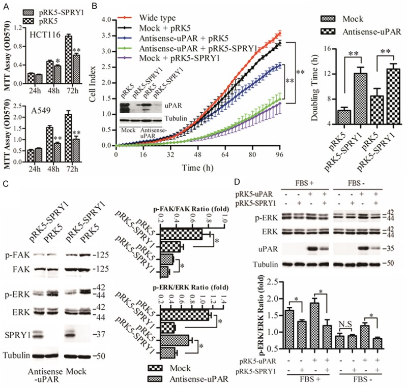
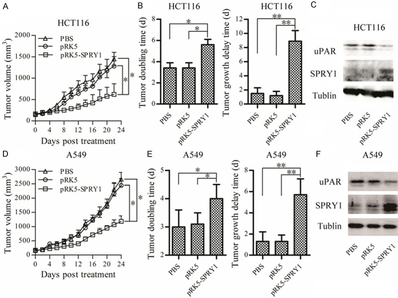
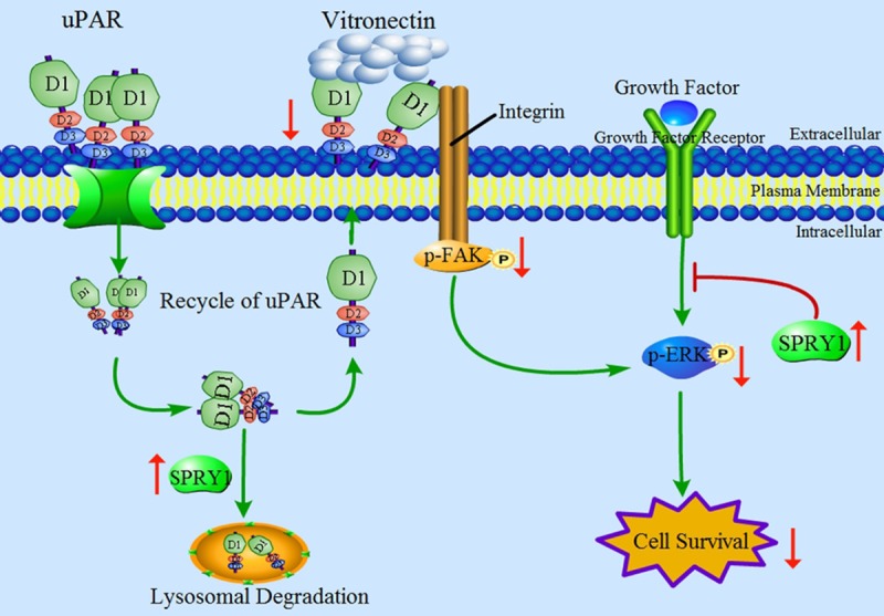
Urokinase plasminogen activator receptor (uPAR) is a GPI anchored cell surface protein that is closely associated with invasion, migration, and metastasis of cancer cells. Many functional extracellular proteins and transmembrane receptors interact with uPAR. However, few studies have examined the association of uPAR with cytoplasm proteins. We previously used yeast two-hybrid screening to isolate several novel uPAR-interacting cytoplasmic proteins, including Sprouty1 (SPRY1), an inhibitor of the (Ras-mitogen-activated protein kinase) MAPK pathway. In this study, we show that SPRY1 interacts with uPAR and directs it toward lysosomal-mediated degradation. Overexpression of SPRY1 decreased the cell surface and cytoplasmic uPAR protein level. Moreover, SPRY1 overexpression augmented uPAR-induced cell adhesion to vitronectin as well as proliferation of cancer cells. Our results also further support the critical role of SPRY1 contribution to tumor growth. In a subcutaneous tumor model, overexpression of SPRY1 in HCT116 or A549 xenograft in athymic nude mice led to great suppression of tumor growth. These results show that SPRY1 may affect tumor cell function through direct interaction with uPAR and promote its lysosomal degradation.

Keywords: SPRY1; adhesion; degradation; proliferation; uPAR.


Figures

Figure 1
Figure 1
Real-time PCR results measuring relative levels of SPRY1 mRNA in the different cell types. Relative SPRY1 mRNA levels in different cell types were determined in relation to the housekeeping gene β-actin. mRNA levels obtained from HT29 cells were set at 1. All other levels were normalized to this value. Data from RT-PCR experiments are shown as means ± SEM of three independent experiments.
Figure 2
Figure 2
Immunofluorescence localization of SPRY1 and uPAR proteins in HeLa cells. Upper panel: Endogenous uPAR (red) and SPRY1 (green) co-localized in the cytoplasm. The cells were stained for SPRY1 using anti-SPRY1 antibody and Alexa Fluor 488-conjugated anti-mouse IgG (green). uPAR was detected by anti-uPAR antibody and Alexa Fluor 594-conjugated anti-rabbit IgG (red). Middle panel: Cells were transfected with pRK5-uPAR-His vector and exogenous uPAR was detected by immunostaining with anti-His antibody (in red). Lower panel: Cells were transfected with pRK5-SPRY1-Flag and exogenous SPRY1 was detected by immunostaining with anti-Flag antibody (in green). The nuclei were stained with DAPI (blue).
Figure 3
Figure 3
SPRY1 promotes uPAR downregulation. A. Western blot analysis of SPRY1 and uPAR protein amounts in total cell lysates from A549, HCT116 and 293-uPAR cells, transfected with pRK5-SPRY1 or pRK5. Tubulin was used as loading control. B. uPAR mRNA levels in A549, HCT116 and 293-uPAR cells (transfected with pRK5-SPRY1 or empty vector) were analyzed by quantitative PCR. β-actin was used for normalization of gene expression. C. Western blot analysis of SPRY1 and uPAR protein amounts in total cell lysates from 293-uPAR cells transfected with increasing amounts of SPRY1. Tubulin was used as loading control. D. Cells were transfected with increasing amounts of pRK5-SPRY1 or pRK5 empty vector, and cytotoxic effects of transfection to the expression of uPAR proteins were assessed. E. 293-uPAR cells transfected with pRK5-SPRY1 were treated with 20 µg/ml CHX and cells were lysed at the indicated times. Stability of uPAR was determined by western blot. Tubulin was used as loading control. F. SPRY1 decreases the stability of uPAR via the proteosome pathway. SPRY1 plasmids were transfected into 293-uPAR cells. Cells were treated with CHX (20 µg/ml), MG132 (20 µM) or E64 (20 µM) for indicated time points, and the cell lysates were evaluated by western blot. Each experiment was independently repeated three times. Tubulin was used as loading control. N.S.: no significant difference.
Figure 4
Figure 4
SPRY1 decrease of uPAR-mediated cell adhesion to vitronectin. (A) Fibronectin, vitronectin or BSA proteins were coated on the cell culture plate and blocked with BSA. HCT116 or A549 cells (transfected with pRK5-SPRY1 or empty vector) were spread on the immobilized proteins for 1 h. After washing, adherent cells were fixed with methanol, and stained with Giemsa stain. The relative numbers of cells were quantified using Safire Fluorescence Absorbance at OD550. The experiments were repeated twice and each column represents the mean of a triple determination. **P < 0.01: significantly different from the indicated control. (B) 293 and 293-uPAR cells were transfected with pRK5-SPRY1 or empty vector, and cell adhesion to fibronectin, vitronectin or BSA were evaluated as in HCT116 or A549 cells. Representative micrographs of the attached cells were examined microscopically. (C) 293-uPAR cells were transfected with pRK5-SPRY1 or empty vector, and 24 h later, the cell surface uPAR was analyzed by flow cytometry. (D) Whole cells in (C) were lysed by ultrasonic disruption and the expression of uPAR was detected by western blot. Tubulin was used as control. Each experiment was independently repeated three times. N.S.: no significant difference.
Figure 5
Figure 5
SPRY1 inhibition of uPAR induced cell proliferation. A. Effects of SPRY1 on the proliferation of HCT116 and A549 cells. Cells transfected with pRK5-SPRY1 or empty vector were seeded into 96-well plates. The cells were cultured for 24-72 h followed by MTT assay (OD570) to quantitate cell growth. Data are shown as mean ± SEM from three independent experiments. *P < 0.05, **P < 0.01 versus vector control. B. Effects of SPRY1 on the proliferation of antisense-uPAR or mock transfected HCT116 stable cell lines. Antisense-uPAR and mock cells were transfected with equal amounts of pRK5-SPRY1 or empty vector for 12 h, and then seeded on Real-Time Cellular Analysis (RTCA) 16 well E-plates with 1×10⁴ cells/well (n = 3). Dynamic monitoring of cell proliferation was performed using the xCELLigence RTCA system for an additional 96 h. Relative cell doubling time from 0 to 48 h was performed using RTCA Software 1.2.1. The results are representative of two independent experiments performed in quadruplicate. *P < 0.05, **P < 0.01 versus indicated control. C. Effects of SPRY1 on the activation status of FAK and ERK in antisense-uPAR or mock cells. Cells were transiently transfected with pRK5-SPRY1 or empty vector for 48 h. ERK and FAK-397 phosphorylation was determined by western blot analysis. The intensities of the bands were quantified using ImageJ software. Data are from three independent experiments; *P < 0.05 versus indicated control. D. Effects of SPRY1 on uPAR-induced ERK activation. HCT116 cells were transfected with the indicated SPRY1 and uPAR expression vectors together or alone, and 36 h later, cells were continuously cultured in DMEM (without or with FBS added) for another 12 h. ERK phosphorylation was determined by western blot analysis. Data are from three independent experiments; *P < 0.05 versus indicated control.
Figure 6
Figure 6
Delivery of SPRY1 to tumor tissues suppressed solid tumor growth. A549 or HCT116 cells were injected into 6-8-week-old BL/6mice. When tumors reached a size of 150 mm3, mice were intratumorally injected with M-PEI complexed with SPRY1 overexpression vector or empty vector. Tumor volume comparison among different groups in nude mice bearing (A) HCT116 carcinomas and (D) A549 carcinomas (mean ± SD, n = 8, *P < 0.05, compared with each corresponding treatment). Tumor doubling time and tumor growth delay time comparison among different groups in nude mice bearing (B) HCT116 carcinomas and (E) A549 carcinomas. The results are expressed as mean ± SD from 8 animals. *P < 0.05, **P < 0.01 compared to control. SPRY1 and uPAR expression in HCT116 derived tumors (C) and A549 derived tumors (F) were determined by western blot analysis.
Figure 7
Figure 7
Schematic model of the proposed mechanism for SPRY1 promotion of lysosomal degradation of uPAR and inhibition of uPAR-mediated cell adhesion and proliferation. First, upregulation of SPRY1 could inhibit growth factor receptor signaling pathway and inhibit Raf/MEK/ERK-induced proliferation. On the other hand, SPRY1 interacts with uPAR to trigger degradation of uPAR via the lysosomal pathway, and then decrease the amount of uPAR recycled back to the cell surface. Cell surface uPAR decrease not only reduces adhesion functions, but also inhibits activation of FAK and ERK, which finally inhibits cell proliferation.

References

    1. Alpízar-Alpízar W, Nielsen BS, Sierra R, Illemann M, Ramírez JA, Arias A, Durán S, Skarstein A, Ovrebo K, Lund LR, Laerum OD. Urokinase plasminogen activator receptor is expressed in invasive cells in gastric carcinomas from high- and low-risk countries. Int J Cancer. 2010;126:405–15. - PubMed
    1. Wei Y, Waltz DA, Rao N, Drummond RJ, Rosenberg S, Chapman HA. Identification of the urokinase receptor as an adhesion receptor for vitronectin. J Biol Chem. 1994;269:32380–8. - PubMed
    1. Bohuslav J, Horejsí V, Hansmann C, Stöckl J, Weidle UH, Majdic O, Bartke I, Knapp W, Stockinger H. Urokinase plasminogen activator receptor, beta 2-integrins, and Src-kinases within a single receptor complex of human monocytes. J Exp Med. 1995;181:1381–90. - PMC - PubMed
    1. Resnati M, Guttinger M, Valcamonica S, Sidenius N, Blasi F, Fazioli F. Proteolytic cleavage of the urokinase receptor substitutes for the agonist-induced chemotactic effect. EMBO J. 1996;15:1572–82. - PMC - PubMed
    1. Nalla AK, Asuthkar S, Bhoopathi P, Gujrati M, Dinh DH, Rao JS. Suppression of uPAR retards radiation-induced invasion and migration mediated by integrin β1/FAK signaling in medulloblastoma. PLoS One. 2010;5:e13006. - PMC - PubMed

LinkOut - more resources