. 2014 Nov 19;4(6):897-906.
eCollection 2014.

MicroRNA-543 acts as an oncogene by targeting PAQR3 in hepatocellular carcinoma

Affiliations

MicroRNA-543 acts as an oncogene by targeting PAQR3 in hepatocellular carcinoma

Lei Yu et al. Am J Cancer Res. .

Abstract

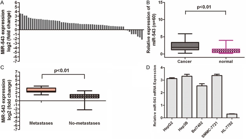
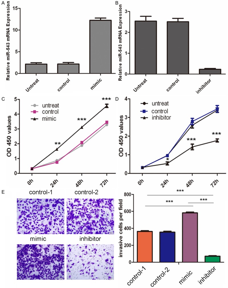
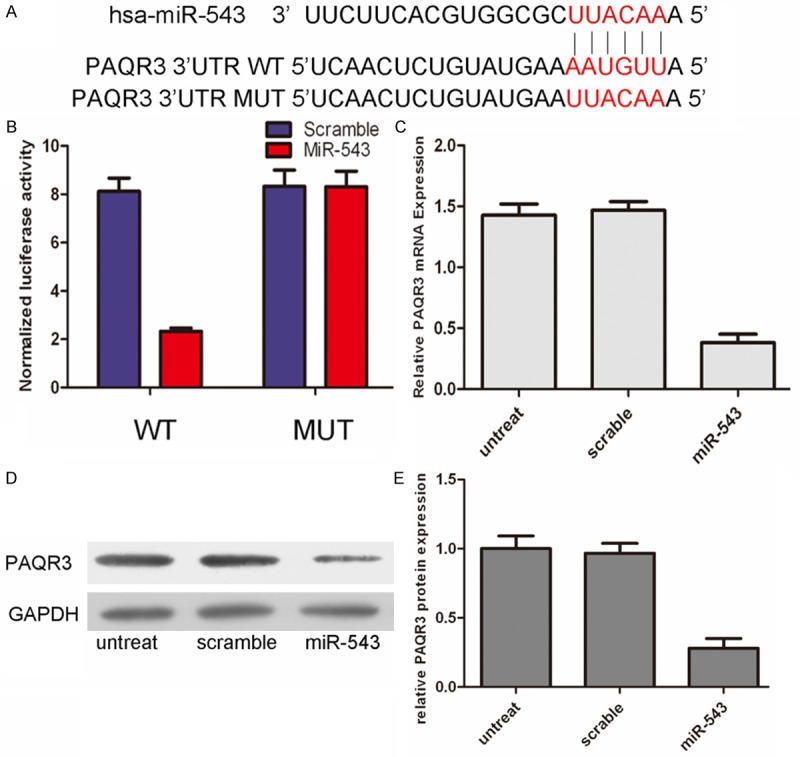
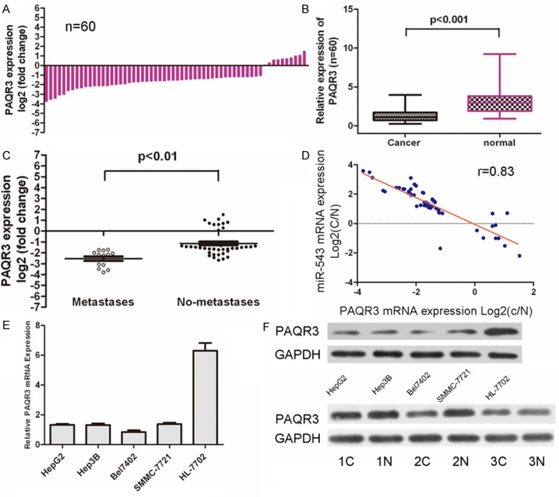
MicroRNAs (miRNAs) are small, non-coding RNAs that can act as oncogenes or tumor suppressor genes in human cancer. Increasing evidences indicate that deregulation of miRNAs contributes to the hepatocarcinogenesis. In this study, we demonstrated that the levels of miR-543 were dramatically increased in clinical hepatocellular carcinoma (HCC) tissues and cell lines. Moreover, forced expression of miR-543 promoted the proliferative and invasive potential of HepG2. We also identified PAQR3 as a direct target gene for miR-543 using a fluorescent reporter assay and western blot. The levels of PAQR3 were dramatically decreased in clinical hepatocellular carcinoma (HCC) tissues and cell lines. The mRNA levels of PAQR3 were inversely correlated with the miR-543 expression level.Thus, our finding provides a new insight into the mechanism of hepatocarcinogenesis, indicating a therapeutic potential of miR-543 in the treatment of HCC.

Keywords: PAQR3; hepatocellular carcinoma; miR-543; oncogene.


Figures

Figure 1
Figure 1
MiR-543 is over-expressed in hepatocellular carcinoma tissues. A. qRT-PCR analysis of miR-543 expression in 60 pairs HCC tissues and their corresponding adjacent nontumorous livers. The expression of miR-543 was normalized to U6 snRNA. B. Relative miR-543 expression levels in HCC tissues and adjacent normal regions; C. Statistical analysis of the association between miRNA level, pM stage (No metastasis and Metastasis, respectively). D. qRT-PCR analysis of miR-543 expression in HCC cells (HepG2, Hep3B, Bel7402, SMMC-7721) and normal hepatocytes (HL-7792 cells).
Figure 2
Figure 2
MiR-543 promotes hepatocellular carcinoma cell growth and invasion. A. qRT-PCR analysis of miR-543 expression after the transfection of miR-543 mimic, or control or no transfection. The expression of miR-543 was normalized to U6 snRNA. B. qRT-PCR analysis of miR-543 expression after the transfection of miR-543 inhibitor, or control or no transfection. The expression of miR-543 was normalized to U6 snRNA. C. The CCK8 assay analysis was used to evaluate the proliferation of HepG2 cells after transfection with the miR-543 mimic, or scramble or no transfection. D. The CCK8 assay analysis was used to evaluate the proliferation of HepG2 cells after transfection with the miR-543 inhibitor, or scramble or no transfection. E. Transwell analysis of the HepG2 cells after treatment with miR-543 mimics, inhibitors or control or no transfection; the invasive cells per field is shown below, *p < 0.05, **p < 0.01, and ***p < 0.001.
Figure 3
Figure 3
PAQR3 was direct target of miR‑543. A. The sequences of miR-543 binding sites within the human PAQR3 3’UTRs and schematic reporter constructs, in this panel, PAQR3-WT represent the reporter constructs containing the entire 3’UTR sequences of PAQR3. PAQR3-MUT represent the reporter constructs containing mutated nucleotides. B. The analysis of the relative luciferase activities of PAQR3-WT, PAQR3-MUT in 293T cells. The error bars are derived from triplicate experiments. C. qRT-PCR analysis of PAQR3 mRNA expression in HepG2 cells after treatment with miRNA mimics or scramble or no transfection. The expression of PAQR3 was normalized to GAPDH. D, E. Western blot analysis of PAQR3 expression in HepG2 cells transfected with miR-543 mimics or scramble or no transfection. GAPDH was also detected as a loading control.
Figure 4
Figure 4
MiR‑543 expression inversely correlated with PAQR3 mRNA in HCC samples. A. qRT-PCR analysis of PAQR3 expression in 60 pairs HCC tissues and their corresponding adjacent nontumorous livers. The expression of PAQR3 was normalized to GAPDH mRNA. B. Relative PAQR3 expression levels in HCC tissues and adjacent normal regions; C. Statistical analysis of the association between PAQR3 level, pM stage (No metastasis and Metastasis, respectively). D. The mRNA expression of PAQR3 was inversed correlation the expression of miR-543 levels in patient samples. E. qRT-PCR analysis of PAQR3 expression in HCC cells (HepG2, Hep3B, Bel7402, SMMC-7721) and normal hepatopcytes (HL-7792 cells). F. Western blot analysis of PAQR3 expression in the HCC cells (HepG2, Hep3B, Bel7402, SMMC-7721) and normal hepatopcytes (HL-7792 cells); and 3 pairs NSCLC tissues and their corresponding adjacent normal lung tissues.

References

    1. Caffarelli E, Filetici P. Epigenetic regulation in cancer development. Front Biosci (Landmark Ed) 2011;16:2682–2694. - PubMed
    1. Rosell R, Cuello M, Cecere F, Santarpia M, Reguart N, Felip E, Taron M. Usefulness of predictive tests for cancer treatment. Bull Cancer. 2006;93:E101–108. - PubMed
    1. Wang PR, Xu M, Toffanin S, Li Y, Llovet JM, Russell DW. Induction of hepatocellular carcinoma by in vivo gene targeting. Proc Natl Acad Sci U S A. 2012;109:11264–11269. - PMC - PubMed
    1. Zucman-Rossi J. Molecular classification of hepatocellular carcinoma. Dig Liver Dis. 2010;42(Suppl 3):S235–241. - PubMed
    1. Banaudha KK, Verma M. The role of microRNAs in the management of liver cancer. Methods Mol Biol. 2012;863:241–251. - PubMed

LinkOut - more resources