Review
. 2014 Dec 3:2:265.
doi: 10.3389/fpubh.2014.00265. eCollection 2014.

Chagas' disease: an emergent urban zoonosis. The caracas valley (Venezuela) as an epidemiological model

Affiliations
Review

Chagas' disease: an emergent urban zoonosis. The caracas valley (Venezuela) as an epidemiological model

Servio Urdaneta-Morales. Front Public Health. .

Abstract

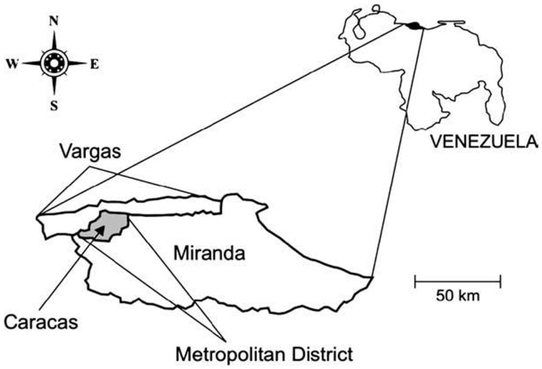
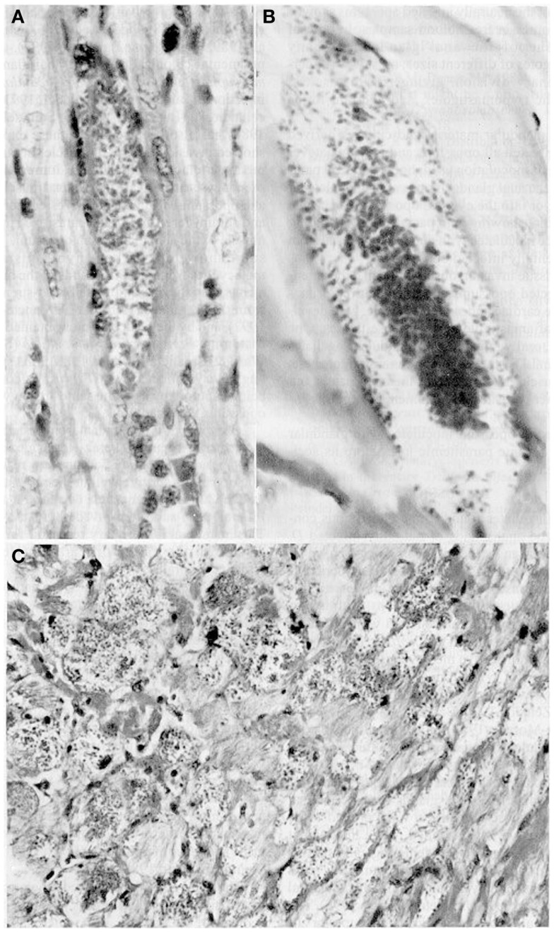
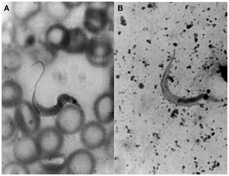
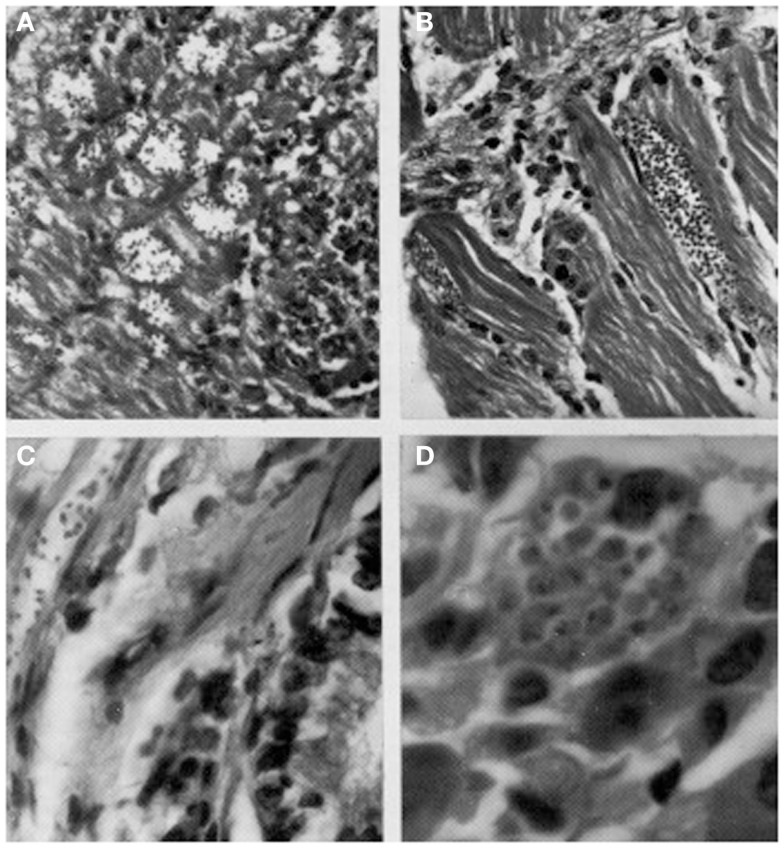
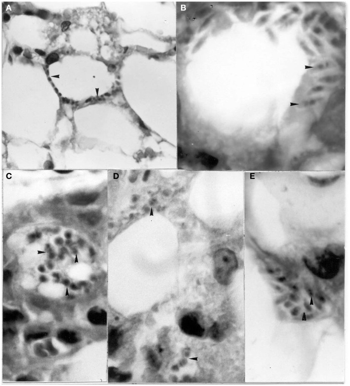
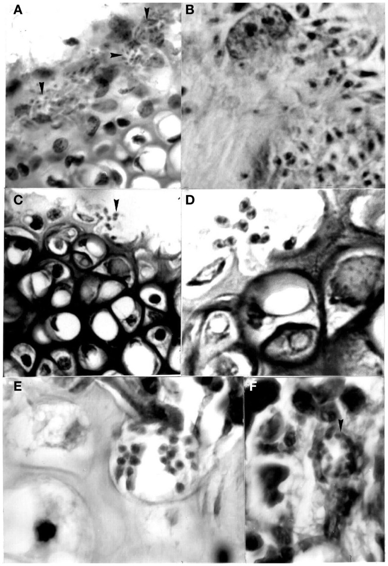
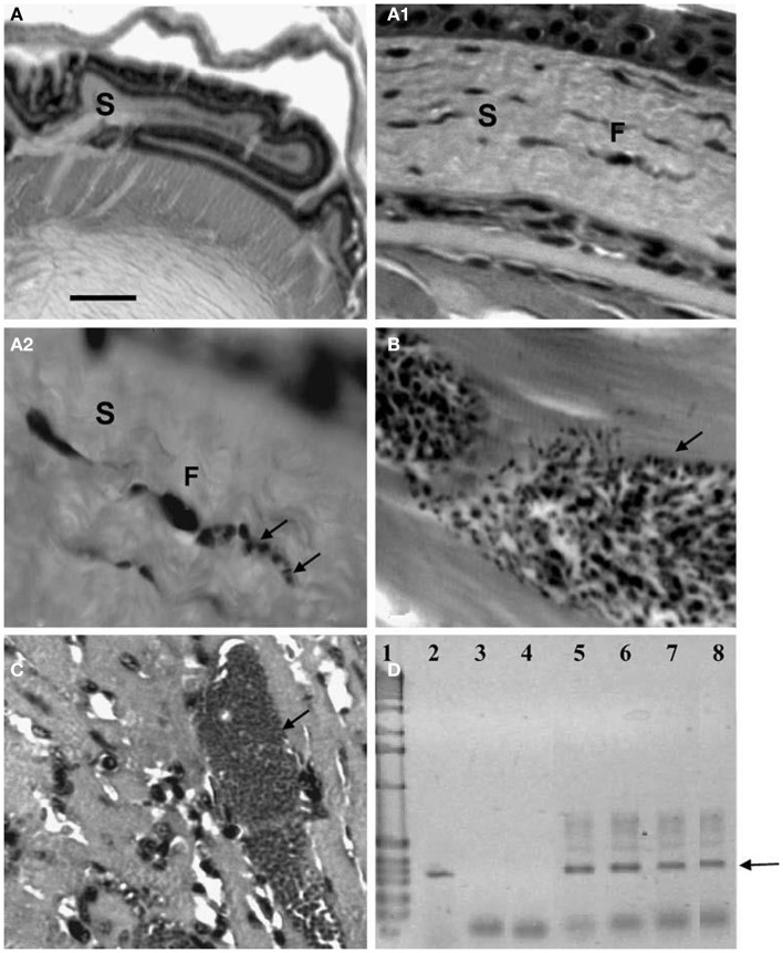
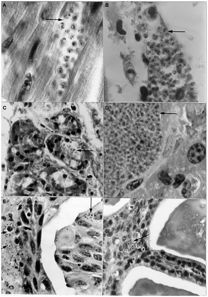
The unprecedented emergence of important public health and veterinary zoonoses is usually a result of exponential population growth and globalization of human activities. I characterized Chagas' disease as an emergent zoonosis in the Caracas Valley (Venezuela) due to the following findings: the presence of reservoirs (Didelphis marsupialis, Rattus rattus) and vectors (Panstrongylus geniculatus, Panstrongylus rufotuberculatus) infected with Trypanosoma cruzi in urbanized or marginalized areas; the elevated contact between P. geniculatus and human beings detected by parasitological and molecular examinations of triatomine feces demonstrated the possibility of transmission risks; a study of outbreaks of urban Chagas' disease reported the first proven case of oral transmission of T. cruzi to human beings; the risk of transmission of glandular metacyclic stages from marsupials by experimental ocular and oral instillation; mice genitalia infected with T. cruzi contaminated blood resulted in the formation of amastigotes very close to the lumen suggesting that there may be a possibility of infection via their release into the urine and thence to the exterior; the ubiquitous histotropism and histopathology of T. cruzi was demonstrated using a mouse model; the presence of experimental T. cruzi pseudocysts in adipose, bone-cartilage, and eye tissue indicated a potential risk for transplants. Socio-sanitary programs that include improvements in housing, vector control, and access to medical treatment, as well as strategies aimed at combating social inequalities, poverty, and underdevelopment should be undertaken in those areas where zoonoses are most prevalent. Disciplines, such as Ecology, Epidemiology, Medical Entomology, Human and Veterinary Medicine, Environmental Studies, Public Health, Social and Political Studies, Immunology, Microbiology, and Pharmacology could all provide important contributions that aim to reduce the occurrence of factors governing the spread of emergent diseases.

Keywords: Caracas Valley (Venezuela); Chagas’ disease; emerging urban zoonosis.


Figures

Figure 1
Figure 1
Map of Venezuela showing the relative location of the area of the Metropolitan District.
Figure 2
Figure 2
Tissue sections showing pseudocysts containing amastigotes (H-E). (A) Heart of Didelphis marsupialis infected orally by glandular material cultured in LIT medium (400×; (B) muscle layer of anal gland of opossum, infected as above (1000×); (C) heart of mouse infected intraperitoneally by glandular material from a naturally infected opossum (400×).
Figure 3
Figure 3
Flagellate stages of Trypanosoma cruzi: (A) stout bloodstream trypomastigote from naturally infected Rattus rattus (Giemsa, 1400×); (B) metacyclic trypomastigote from feces of Rhodnius prolixus used for xenodiagnosis of naturally infected R. rattus (Giemsa, 1400×).
Figure 4
Figure 4
Histological sections showing pseudocysts of Trypanosoma cruzi with amastigotes in (A) cardiac tissue of naturally infected Rattus rattus (H-E; 560×); (B) skeletal muscle of naturally infected R. rattus (H-E, 560×); (C) smooth muscle fiber from the colon of an experimentally infected mouse (H-E, 960×); (D) acinar cell of pancreas of experimentally infected mouse (H-E, 1400×).
Figure 5
Figure 5
Amastigotes and intermediate stages (arrows) of Trypanosoma cruzi in (A,B) perifery cytoplasm of an adipocye; (C) cytoplasm of immature adipocite (preadipocite); (D) intercellular substance in connective adipose tissue; (E) parasitized macrophage located between uninfected adipocytes (H-E; 1400×).
Figure 6
Figure 6
Pseudocysts with stages (arrows) of Trypanosoma cruzi in sternum of mice experimentally infected with different isolates. (A) Perichondrium with nests showing amastigotes and intermediate stages in chondroblasts (400×; HE); (B) same stages in stroma of perichondrium and in the cytoplasm of a macrophage (1000×; HE); (C) and (D) two broken chondrocytes with several amastigotes and one flagellated stage (400× and 1000×, respectively; HE); (E) several amastigotes in a osteocyte (1000×; HE); (F) pseudocyst with amastigotes in the bone marrow (1000×; HE).
Figure 7
Figure 7
Histological and molecular parasitism in NMRI mice experimentally infected with different isolates and strains of T. cruzi. (A – A2). Sequence of microphotographs with amplification of a nest of amastigotes in a fibroblast (F; arrows; 40×, 400×, and 1000×, respectively) of corneal stroma (S); (B) trypomastigote nest in thigh skeletal muscle (arrow); (C) amastigote nest in heart muscle (arrow); (D) amplification of the 330-bp fragment from the conserved regions of kDNA (arrow) extracted from ocular tissues of experimentally infected NMRI mice in 2.5% agarose gel electrophoresis (ethidium bromide stain): Lane 1 1-kb ladder molecular marker (Gibco BRL Life Technologies), lane 2 nude T. cruzi DNA, lane 3 negative PCR control, lane 4 MRAT/VE/1996/CO22 isolate, lane 5 MHOM/BR/1950/Y strain, lane 6 MHOM/VE/1970/EP isolate, lane 7 MDID/BR/1999/M1 isolate, and lane 8 MDID/VE/1995/CO79 isolate.
Figure 8
Figure 8
Histological sections from albino mice intravaginally instilled with a strain of Trypanosoma cruzi from Rattus rattus, and scrotally inoculated with a strain from a human patient, showing nests (arrows) with amastigotes in (A) cardiac tissue; (B) skeletal muscle; (C) pancreas (acinus); (D) liver; (E) urinary bladder (epithelium very close to the lumen and lamina propria); (F) seminal vesicle (mucosa close to the lumen). [(A,B,D): l400×; (C,E,F): 950×; H-E].

References

    1. Pinto Dias JC. Tendencias sociales de la enfermedad de Chagas para las próximas décadas. Sal Colect (2012) 8(Suppl 1):1218–9.
    1. Galvão C, Carcavallo R, Rocha D, Jurberg J. A checklist of the current valid species of the subfamily Triatominae jeannnel, 1919 (Hemiptera, Reduviidae) and their geographical distribution, with nomenclatural and taxonomic notes. Zootaxa (2003) 202:1–36.
    1. Coura JR. Chagas disease: what is known and what is needed-a background article. Mem Inst Oswaldo Cruz (2007) 102(Suppl 1):113–22. 10.1590/S0074-02762007000900018 - DOI - PubMed
    1. Teixeira AR, Monteiro PS, Rebelo JM, Argañaraz ER, Vieira D, Lauria-Pires L, et al. Emerging Chagas disease: trophic network and cycle of transmission of Trypanosoma cruzi from palm trees in the Amazon. Emerg Infect Dis (2001) 7:100–12. 10.3201/eid0701.070100 - DOI - PMC - PubMed
    1. Briceño-León R. La enfermedad de Chagas en las Américas: una perspectiva de ecosalud. Cad Saúde Pública (2009) 25(Suppl 1):S71–82. 10.1590/S0102-311X2009001300007 - DOI - PubMed

LinkOut - more resources