. 2015 Jan;25(1):35-43.
doi: 10.1111/bpa.12219.

Age-associated white matter lesions: the MRC Cognitive Function and Ageing Study

Affiliations

Age-associated white matter lesions: the MRC Cognitive Function and Ageing Study

Stephen B Wharton et al. Brain Pathol. 2015 Jan.

Abstract

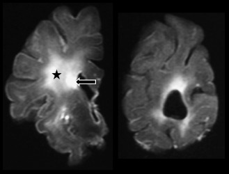
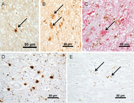
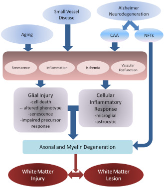
Cerebral white matter lesions (WML) are common in the aging brain and are associated with dementia and depression. They are associated with vascular risk factors and small vessel disease, suggesting an ischemic origin, but recent pathology studies suggest a more complex pathogenesis. Studies using samples from the population-representative Medical Research Council Cognitive Function and Ageing Study neuropathology cohort used post-mortem magnetic resonance imaging to identify WML for further study. Expression of hypoxia-related molecules and other injury and protective cellular pathways in candidate immunohistochemical and gene expression microarray studies support a role for hypoxia/ischemia. However, these approaches also suggest that immune activation, blood-brain barrier dysfunction, altered cell metabolic pathways and glial cell injury contribute to pathogenesis. These abnormalities are not confined to WML, but are also found in apparently normal white matter in brains with lesions, suggesting a field effect of white matter abnormality within which lesions arise. WML are an active pathology with a complex pathogenesis that may potentially offer a number of primary and secondary intervention targets.

Keywords: aging; blood-brain barrier; dementia; small vessel disease; vascular; white matter lesions.


Figures

Figure 1
Figure 1
Magnetic resonance imaging scan of fixed brain slices. Left: high signal in the white matter identifies a periventricular lesion (arrow) and a deep white matter lesion (star). Right: slice at occipital level showing periventricular areas of high signal.
Figure 2
Figure 2
A. Immunohistochemistry for +13 isoform of microtubule‐associated protein‐2 identifies a process‐bearing oligodendrocyte precursor cells (arrow). B. Clasmatodendritic astrocytes labeled for fibrinogen (arrows). C. Double‐immunohistochemistry for glial fibrillary acidic protein (GFAP) (red) and CD68 (brown). The clasmatodendritic astrocyte labels for GFAP (arrow), but not CD68, antibody to which shows granular labeling of microglial processes. D. Amoeboid microglia in a deep subcortical white matter lesions. E. Ramified microglial labeled for major histocompatibility complex class II in a periventricular lesion (arrows). Magnification bars all 50 μm.
Figure 3
Figure 3
Possible pathogenic mechanisms for white matter damage in the context of vascular and Alzheimer‐related pathologies. Factors such as aging and small vessel disease may result in glial injury and inflammation, acting via a number of pathogenic mechanisms, including cell damage and senescence, inflammation, ischemia and vascular dysfunction, which may include blood–brain barrier changes and altered drainage. Glial injury and inflammation may then damage myelin and axons, resulting in the myelin pallor observed histologically. Alzheimer's may also act through small vessel ischemia related to cerebral amyloid angiopathy (CAA), while neurodegeneration related to neurofibrillary tangle (NFT) formation may directly cause axonal damage and secondary myelin loss. Although shown with arrows, mechanisms are interactive and processes may go in both directions and produce feed‐forward cycles. Further, different antecedents may cause white matter pallor and dysfunction via different routes and predilection to affect either myelin or axon primarily. These differing routes to white matter injury and lesion formation require definition.

References

    1. Akiguchi I, Tomimoto H, Suenaga T, Wakita H, Budka H (1997) Alterations in glia and axons in the brains of Binswangers disease patients. Stroke 28:1423–1429. - PubMed
    1. Bakker S, de Leeuw F, de Groot C, Hofman A, Koudstaal P, Breteler M (1999) Cerebral vasomotor reactivity and cerebral white matter lesions in the elderly. Neurology 52:578–583. - PubMed
    1. Barber R, Scheltens P, Gholkar A, Ballard C, McKeith I, Ince P et al (1999) White matter lesions on magnetic resonance imaging in dementia with Lewy bodies, Alzheimer's disease, vascular dementia, and normal aging. J Neurol Neurosurg Psychiatr 67:66–72. - PMC - PubMed
    1. Bell R, Winkler E, Singh I, Sagare A, Deane R, Wu Z et al (2012) Apolipoprotein E controls cerebrovascular integrity via cyclophilin A. Nature 485:512–615. - PMC - PubMed
    1. Boche D, Perry V, Nicoll J (2013) Activation patterns of microglia and their identification in the human brain. Neuropathol Appl Neurobiol 39:3–18. - PubMed

MeSH terms