Review
. 2015 Jan;25(1):63-78.
doi: 10.1111/bpa.12218.

White matter changes in dementia: role of impaired drainage of interstitial fluid

Affiliations
Review

White matter changes in dementia: role of impaired drainage of interstitial fluid

Roy O Weller et al. Brain Pathol. 2015 Jan.

Abstract

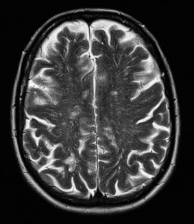
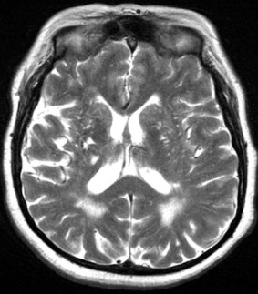
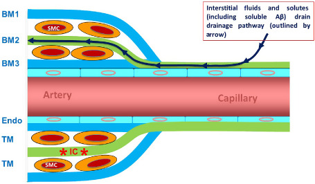
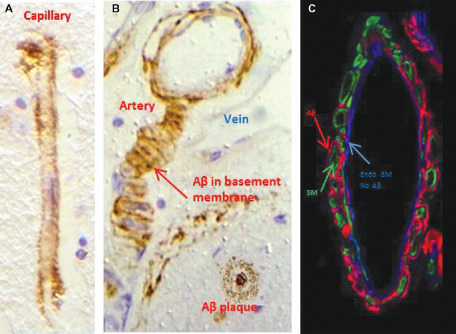
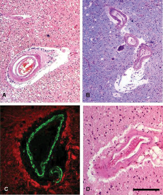
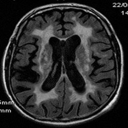
White matter abnormalities on magnetic resonance imaging (MRI) are associated with dementia and include white matter hyperintensities (WMH; also termed leukoaraiosis) and visible perivascular spaces (PVS). We review the potential role of impaired drainage of interstitial fluid in the pathogenesis of WMH and PVS. Whereas the volume of extracellular space in the grey matter is tightly controlled, fluid accumulates and expands the extracellular spaces of the white matter in acute hydrocephalus, vasogenic edema and WMH. Although there are no conventional lymphatic vessels in the brain, there is very effective lymphatic drainage for fluid and solutes along restricted pathways in the basement membranes of cerebral capillaries and arteries in young individuals. Lymphatic drainage of the brain is impaired with age and in association with apolipoprotein E ε4, risk factors for Alzheimer's disease and cerebral amyloid angiopathy (CAA). Deposition of proteins in the lymphatic drainage pathways in the walls of cerebral arteries with age is recognized as protein elimination failure angiopathy (PEFA), as in CAA and cerebral autosomal dominant arteriopathy and leukoencephalopathy (CADASIL). Facilitating perivascular lymphatic drainage from the aging brain may play a significant role in the prevention of CAA, WMH and Alzheimer's disease and may enhance the efficacy of immunotherapy for Alzheimer's disease.

Keywords: Alzheimer's disease; CADASIL; amyloid-β; cerebral amyloid angiopathy; perivascular drainage; perivascular spaces; protein elimination failure angiopathy (PEFA); white matter hyperintensities.


Figures

Figure 1
Figure 1
T2‐weighted axial image showing MRI‐visible PVS in the hemisphere white matter in a patient with CAA‐related ICH.
Figure 2
Figure 2
T2‐weighted MRI axial image showing confluent WMH in association with extensive deep (basal ganglia) MRI‐visible perivascular spaces and lacunar infarcts.
Figure 3
Figure 3
Perivascular drainage. ISF and solutes, including Aβ, diffuse through the extracellular spaces of the brain and then drain out of the brain along basement membranes (BM) in the walls of capillaries and arteries. The drainage pathway is along the BM colored green: that is capillary endothelial BM and the BM of smooth muscle cells (SMC) in the mid‐zone of the tunica media (TM) of the artery (BM2). In the artery wall, BM on the outer aspect (BM1) and on the endothelial aspect (BM3) is not part of the drainage pathway. With reference to Figure 8, immune complexes (*IC*) are trapped within the BM2 pathway and obstruct perivascular drainage. ENDO, endothelium.
Figure 4
Figure 4
Cerebral amyloid angiopathy. Aβ (brown) is deposited in basement membranes in the walls of capillaries (A) and arteries (B,C). Although the basement membranes of smooth muscle cells (SM) in the tunica media contain Aβ (see figures B and C), the endothelial basement membrane (Endo BM) is almost devoid of Aβ. Immunocytochemistry for Aβ (brown or red) and smooth muscle actin (green). Collagen IV in basement membrane in (C) is blue.
Figure 5
Figure 5
Leptomeningeal arteries from wild‐type and APOE ε4 mice following intracerebral injection of fluorescent Aβ (green). Within 5 minutes of the injection, Aβ was observed along basement membranes, colocalizing (pink) with collagen IV (blue) in young, 3‐month‐old wild‐type mice in (A). Aggregation of Aβ is seen as early as 3 months in the APOE4 mice (arrow in B). Aβ continues to drain along basement membranes of aged wild‐type mice (C), but appears as large deposits in 16 months in the APOE ε4 mice (D, arrows). Scale bars = 10 μm in A; 25 μm in B, C and D. Reproduced from reference 50.
Figure 6
Figure 6
Proposed mechanism for CAA‐induced dilatation of perivascular spaces in the white matter. Branches of the leptomeningeal arteries penetrate the cortex (often without branching) to supply the subcortical white matter. CAA in the walls of the leptomeningeal arteries (yellow banding) obstructs the drainage of interstitial fluid and solutes resulting in retention of fluid in dilated perivascular spaces around arteries in the white matter.
Figure 7
Figure 7
Schematic diagram of the temporal sequence of immunization and injection experiments. Wild‐type BALB/c mice were immunized with ovalbumin (OVA) and then injected with OVA in the striatum at different time points. A soluble fixable fluorescent dextran was then injected intracerebrally, 5 minutes or 24 h or 7 days post‐immunization. Reproduced from reference 17 Original Publisher: BioMed Central.
Figure 8
Figure 8
Immune complexes in basement membranes in the walls of cerebral capillaries and arteries. Active immunization with ovalbumin (OVA) was followed by intracerebral challenge with OVA and left in situ for 24 h. The figure shows a longitudinal section of an artery in the striatum at 24 h after injection of antigen. There is colocalization (yellow) of complement (green) with laminin (red) in the basement membranes surrounding smooth muscle cells in the tunica media. Scale bar = 60 μm. Reproduced from reference 17 Original Publisher: BioMed Central.
Figure 9
Figure 9
White matter changes and dilated PVS in non‐amyloid microangiopathies (eg, CADASIL) and CAA. Lobar or intracerebral hemorrhages occur in CAA (unlike in CADASIL) whereas white matter disease and microbleeds (cortical and subcortical) are common to both disorders. Examination of the white matter reveals vascular degenerative processes progressing from loss of smooth muscle cells, wall thickening, fibrinoid necrosis to hyalinosis. Dilated PVS occur because of arterial vascular degeneration and impaired perivascular drainage of fluid and solutes in both CADASIL (A and C) and CAA (B and D). Dilated PVS in the white matter is consistently observed to a greater extent in CADASIL compared with CAA. Sections were stained with Hematoxylin and Eosin (H&E) (A, B, D) and double immunofluorescence (C). In C, the wall of an arteriole is labeled with smooth muscle α‐actin (green) with glial fibrillary acidic protein (GFAP)‐labeled perivascular astrocytic processes (red). Scale bars = 70 μm in A and B; 150 μm in C and D.
Figure 10
Figure 10
WMH in CADASIL. FLAIR MRI scan shows the extent of confluent white matter changes in a 56‐year‐old CADASIL subject carrying the p.Arg182Cys mutation.

References

    1. Abbott NJ (2004) Evidence for bulk flow of brain interstitial fluid: significance for physiology and pathology. Neurochem Int 45:545–552. - PubMed
    1. Arbel‐Ornath M, Hudry E, Eikermann‐Haerter K, Hou S, Gregory JL, Zhao L et al (2013) Interstitial fluid drainage is impaired in ischemic stroke and Alzheimer's disease mouse models. Acta Neuropathol 126:353–364. - PMC - PubMed
    1. Arvanitakis Z, Leurgans SE, Wang Z, Wilson RS, Bennett DA, Schneider JA (2011) Cerebral amyloid angiopathy pathology and cognitive domains in older persons. Ann Neurol 69:320–327. - PMC - PubMed
    1. Asuni AA, Boutajangout A, Scholtzova H, Knudsen E, Li YS, Quartermain D et al (2006) Vaccination of Alzheimer's model mice with Abeta derivative in alum adjuvant reduces Abeta burden without microhemorrhages. Eur J Neurosci 24:2530–2542. - PMC - PubMed
    1. Barua NU, Bienemann AS, Hesketh S, Wyatt MJ, Castrique E, Love S, Gill SS (2012) Intrastriatal convection‐enhanced delivery results in widespread perivascular distribution in a pre‐clinical model. Fluids Barriers CNS 9:2. - PMC - PubMed

Publication types