Review
. 2015 Jan;25(1):79-98.
doi: 10.1111/bpa.12220.

Cognitive impairment in multiple sclerosis: clinical, radiologic and pathologic insights

Affiliations
Review

Cognitive impairment in multiple sclerosis: clinical, radiologic and pathologic insights

Gabriele C DeLuca et al. Brain Pathol. 2015 Jan.

Abstract

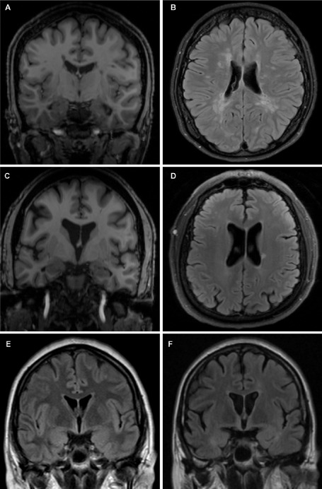
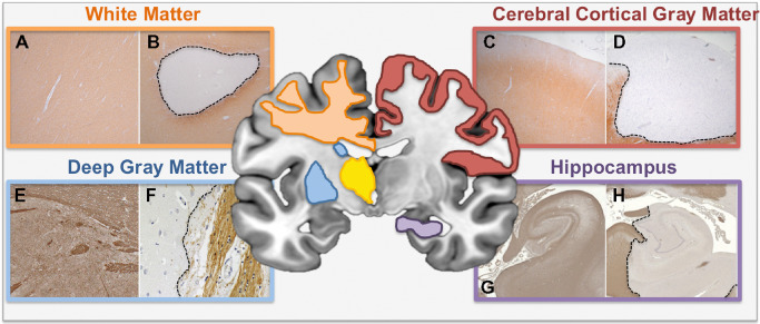
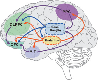
Cognitive impairment is a common and debilitating feature of multiple sclerosis (MS) that has only recent gained considerable attention. Clinical neuropsychological studies have made apparent the multifaceted nature of cognitive troubles often encountered in MS and continue to broaden our understanding of its complexity. Radiographic studies have started to decipher the neuroanatomic substrate of MS-related cognitive impairment and have shed light onto its pathogenesis. Where radiographic studies have been limited by inadequate resolution or non-specificity, pathological studies have come to the fore. This review aims to provide an overview of the nature of cognitive impairment typically seen in MS and to explore the literature on imaging and pathological studies relevant to its evolution. In particular, the relative contributions of gray (i.e., cerebral cortex, hippocampus, thalamus and basal ganglia) and white matter to MS-related cognitive impairment will be discussed and the importance of interconnectivity between structures highlighted. The pressing need for longitudinal studies combining standardized neuropsychometric, paraclinical and radiographic outcomes obtained during life with post-mortem tissue analysis after death is presented.

Keywords: cognitive impairment; gray matter; imaging; multiple sclerosis; pathology; white matter.


Figures

Figure 1
Figure 1
Cortical atrophy, lesion burden and cognitive impairment in MS. Coronal (A, C, E, F) and axial (B, D) views of MRI brain scans from RRMS patients without cognitive impairment (patient 1—A, B; patient 2—C, D) and with cognitive decline (patient 3—E, F). Patient 1 demonstrates relatively preserved brain parenchymal volume (A) despite moderately significant WM lesion burden (B). Conversely, patient 2 has evidence of significant globalized cerebral atrophy (C) in the absence of a significant WM lesion load (D). Patient 3 shows radiographic evidence of progressively worsening global cerebral atrophy over 18 months despite disease‐modifying therapy and relative stability of WM lesion burden. These images highlight the complex relationships between GM and WM pathology and cognitive impairment in MS. A, C. T1‐weighted sequences; B, D, E, F. T2‐FLAIR sequence.
Figure 2
Figure 2
White and gray matter pathology in MS. Schematic diagram outlining the anatomical regions of WM and GM pathology covered in this review (white matter—orange box; deep gray matter—blue box; cerebral cortical GM—red box; hippocampus—purple box). Myelin immunolabeled sections illustrate normal or normal appearing WM/GM (left panel within each colored box—A, C, E, G) or demyelination (right panel in each colored box—B, D, F, H). Specifically, well‐demarcated subcortical WM (B), cerebral cortical subpial (D), caudate GM (F) and hippocampal mixed GM/WM (H) lesions are presented alongside MS subcortical NAWM (A), MS cerebral cortical NAGM (C), non‐neurologic control caudate nucleus (E) and non‐neurologic control hippocampus (H), respectively. A–D. Proteolipid protein immunolabeling. E–H. Myelin basic protein immunolabeling. E, F. Adapted from 208. G, H. Adapted from 166 and used with permission.
Figure 3
Figure 3
Connectivity in the brain and cognitive function. Cognitive function relies on complex interconnections between cortical (cerebral cortex and hippocampus) and deep gray matter structures (eg, thalamus and basal ganglia), with adjoining white matter tracts being important conduits. In the cortex, pyramidal neurons connect cortical structures (cortico‐cortical) or project to subcortical areas to form functionally distinct and temporally coordinated networks (purple lines). Various cortical regions are particularly relevant for cognition, including DLPFC (eg, working memory, planning, and cognitive flexibility), OFC (eg, decision making and adaptive learning), PPC (eg, spatial memory and attention) and AIT (eg, semantic memory) to name a few. The thalamus is a highly integrated structure with connections to several cerebral cortical and subcortical structures important for cognitive function (orange lines). The basal ganglia receive inputs from the intralaminar nuclei of the thalamus and several cortical regions, including frontal, inferotemporal and posterior parietal cortex and form several cortico‐basal ganglionic loops important for cognitive function, including executive function, rule‐based learning and working memory (blue lines). The hippocampal formation (not shown) forms a circuit with neocortical and subcortical structures and subserves diverse functions critical for cognition, including memory formation, maintenance and retrieval being key elements. Although shown with arrows, connections may be in both directions and produce feedforward cycles. AIT = anterior inferior temporal cortex; DLPFC = dorsolateral prefrontal cortex; OFC = orbitofrontal cortex; PPC = posterior parietal cortex.

References

    1. Aarsland D, Karlsen K (1999) Neuropsychiatric aspects of Parkinson's disease. Curr Psychiatry Rep 1:61–68. - PubMed
    1. Achiron A, Chapman J, Magalashvili D, Dolev M, Lavie M, Bercovich E et al (2013) Modeling of cognitive impairment by disease duration in multiple sclerosis: a cross‐sectional study. PLoS One 8:e71058. - PMC - PubMed
    1. Achiron A, Polliack M, Rao SM, Barak Y, Lavie M, Appelboim N, Harel Y (2005) Cognitive patterns and progression in multiple sclerosis: construction and validation of percentile curves. J Neurol Neurosurg Psychiatry 76:744–749. - PMC - PubMed
    1. Ajami B, Bennett JL, Krieger C, McNagny KM, Rossi FM (2011) Infiltrating monocytes trigger EAE progression, but do not contribute to the resident microglia pool. Nat Neurosci 14:1142–1149. - PubMed
    1. Albert M, Antel J, Bruck W, Stadelmann C (2007) Extensive cortical remyelination in patients with chronic multiple sclerosis. Brain Pathol 17:129–138. - PMC - PubMed

Publication types