. 2014 Dec 17;84(6):1213-25.
doi: 10.1016/j.neuron.2014.12.010.

Antisense proline-arginine RAN dipeptides linked to C9ORF72-ALS/FTD form toxic nuclear aggregates that initiate in vitro and in vivo neuronal death

Affiliations

Antisense proline-arginine RAN dipeptides linked to C9ORF72-ALS/FTD form toxic nuclear aggregates that initiate in vitro and in vivo neuronal death

Xinmei Wen et al. Neuron. .

Abstract

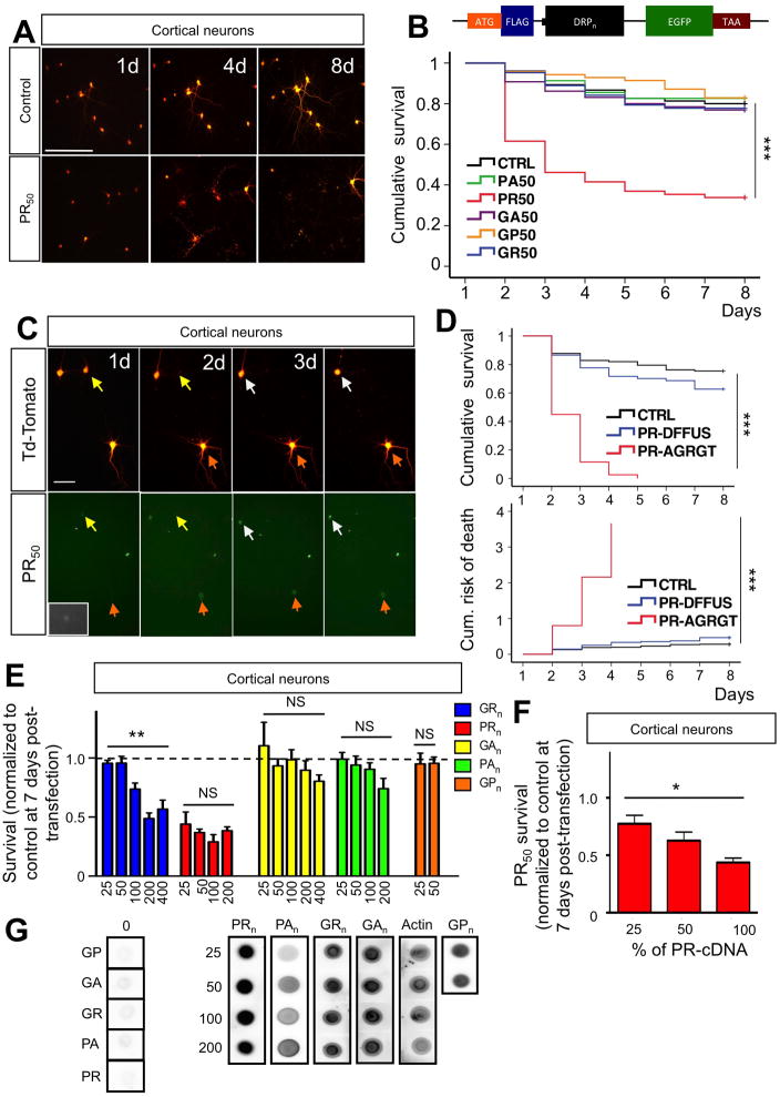
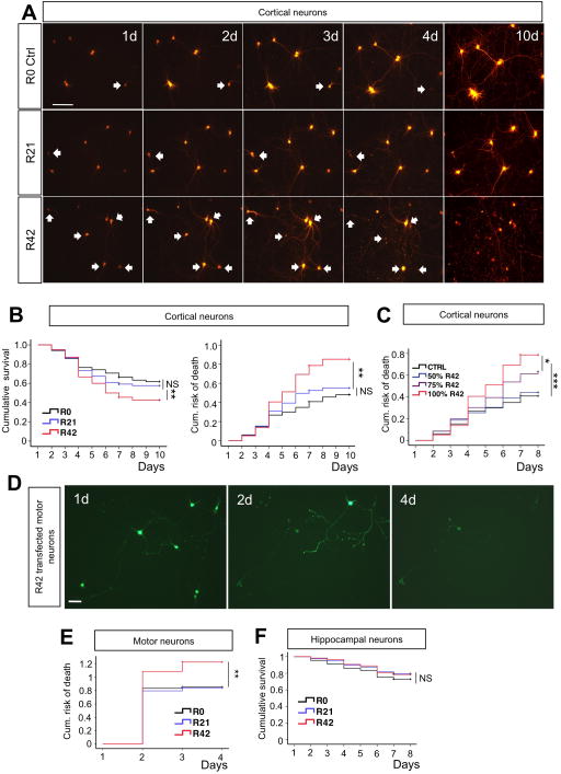
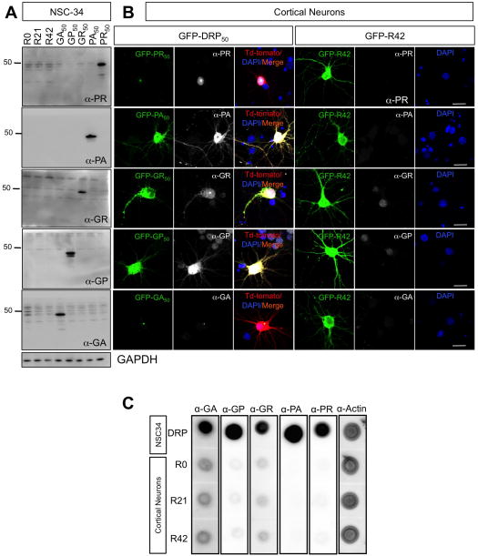
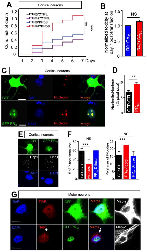
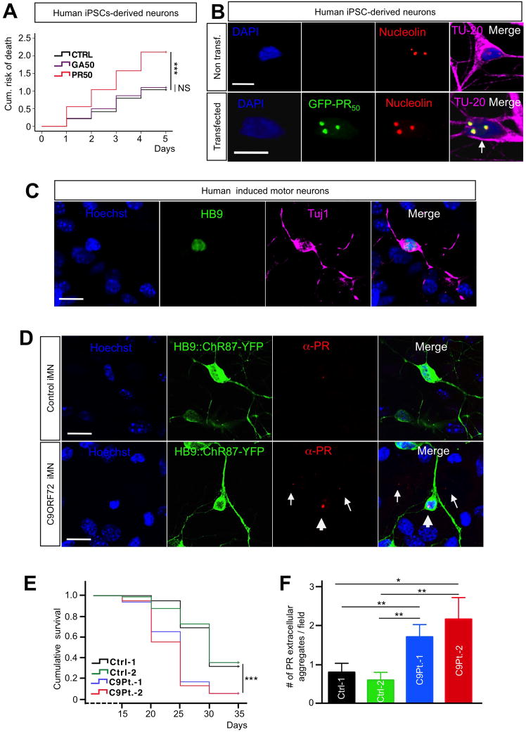
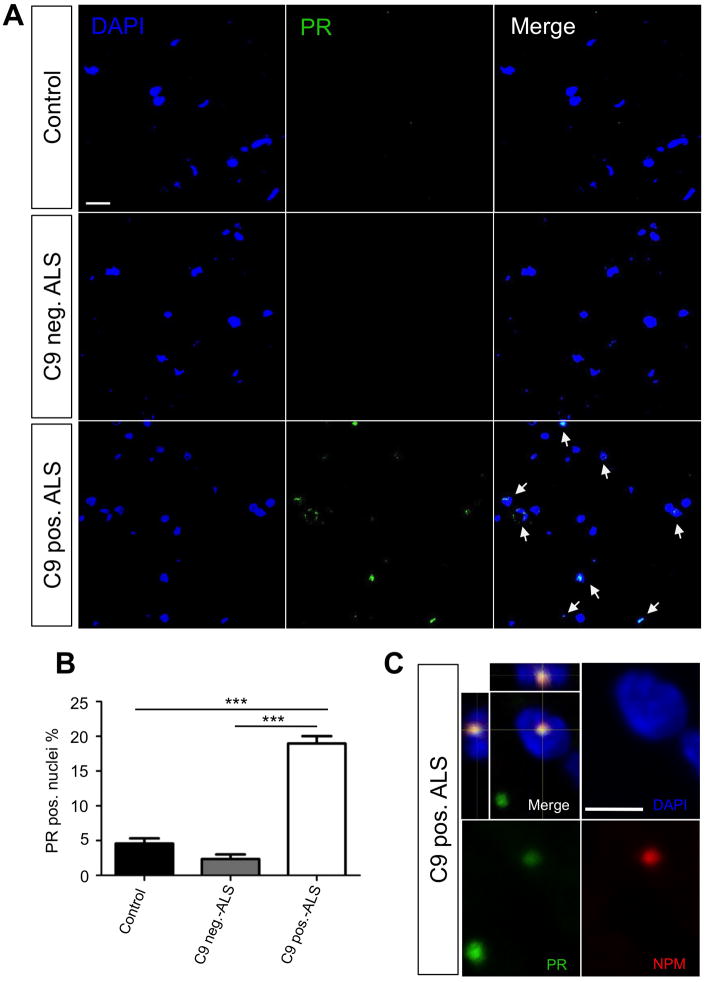
Expanded GGGGCC (G4C2) nucleotide repeats within the C9ORF72 gene are the most common genetic mutation associated with both amyotrophic lateral sclerosis (ALS) and frontotemporal dementia (FTD). Sense and antisense transcripts of these expansions are translated to form five dipeptide repeat proteins (DRPs). We employed primary cortical and motor neuron cultures, live-cell imaging, and transgenic fly models and found that the arginine-rich dipeptides, in particular Proline-Arginine (PR), are potently neurotoxic. Factors that anticipated their neurotoxicity included aggregation in nucleoli, decreased number of processing bodies, and stress granule formation, implying global translational dysregulation as path accountable for toxicity. Nuclear PR aggregates were also found in human induced motor neurons and postmortem spinal cord tissues from C9ORF72 ALS and ALS/FTD patients. Intronic G4C2 transcripts, but not loss of C9ORF72 protein, are also toxic to motor and cortical neurons. Interestingly, G4C2 transcript-mediated neurotoxicity synergizes with that of PR aggregates, suggesting convergence of mechanisms.


Figures

Figure 1
Figure 1. Neurotoxicity of C9RAN poly-dipeptides
(A) Representative live-cell images of cortical neurons co-transfected at DIV10 with control-GFP + Td-tomato plasmids (4:1) or PR50-GFP + Td-Tomato (4:1). The same neurons were imaged for 9 days (8 days post-transfection) at 24-hour intervals. Calibration bar is 100 μm. (B) Kaplan-Meier survival analysis of cortical neurons co-transfected with constructs encoding different DRPs + Td-Tomato. At least 80 neurons were followed/group; n=5 experiments; ***P<0.001. The insert shows the schematic of the construct. The sequence of the insert is the following: ATG-DYKDDK-KLGR-DPR50-GYRARIHYSSVVEFM-EGFP-TAA. The insert was subcloned into pcDNA3.1 vector and its expression driven by the CMV promoter. The DRP sequence has been constructed with a randomized codon strategy. (C) PR50 aggregates formation preceded neuronal death. Representative images show a group of cortical neurons expressing both PR50 (GFP tagged) and Td-tomato. Neurons in which PR50 formed aggregates underwent cell death within 24–36 hours (yellow arrows, day 1–2; white arrows, day 3–4). Conversely, neurons in which PR50 stayed in a diffused, not aggregated pattern (orange arrow and insets) outlived the neurons with PR aggregates. Calibration bar is 50 μm. (D) Cortical neurons transfected with PR50 that formed aggregates exhibited decreased survival and increased risk of death compared to those that didn’t form detectable PR50 aggregates (***p<0.001). PR50 transfected neurons with no detectable aggregates showed no difference in survival and risk of death compared to control transfected neurons (P = 0.08). (E) Length-dependent toxicity of DRPs in cortical neurons. Poly-GR displayed clear length-dependent toxicity (**P<0.01; one-way ANOVA), whereas the effect of poly-PR already peaked at 25 repeats, displaying no significant length-dependency (one-way ANOVA). (F) Cortical neuron toxicity of PR50 diminished as amount of transfected cDNA was reduced suggesting a dose-dependency in the effect. Maximum amount of cDNA-PR50 transfected was 0.8 μg/well (100%). Total amount of DNA transfected was kept constant across groups (1 μg/well). *P<0.05, **P<0.01, ***P<0.001. (G) NSC34 cells were transfected with indicated constrcuts. Expression levels of different lengths of DRP constructs were assessed using dot blots. Actin was used as a loading control. Comparable expression levels were observed for DRPs of different lengths.
Figure 2
Figure 2. Intronic G4C2 repeats are directly toxic to primary neurons
(A) Representative time-lapse images of cortical neurons co-transfected with Td-Tomato and R0, R21 or R42 constructs (1:4 ratio; 1 μg cDNA/well total). Neurons that went on to die are pointed by arrows. More death events were observed in neurons transfected with R42. Calibration bar is 100 μm. (B, C) Kaplan-Meier survival and Cox proportional hazards analysis of cortical neurons transfected with R0, R21 or R42 showed that expanded intronic G4C2 repeats caused cell death and significantly increased the risk of death. Neurotoxicity of G4C2 decreased with reduced amount of R42 cDNA transfected into neurons, suggesting dose-dependency. Note that 50% of R42 cDNA (or 0.4 μg) is not sufficient to cause toxicity. Total amount DNA used in transfection remained the same across groups. At least 80 neurons were followed/group; n=3 experiments. *P<0.05, **P<0.01, ***P<0.001. (D) Representative live-cell images based on GFP fluorescence at day 1, 2 and 4 post-transfection of R42 construct in motor neurons show clear motor neuron loss. Calibration bar is 50 μm. (E) Risk-of-death analysis of motor neurons transfected at DIV5 with R0–42 constructs. At least 40 motor neurons are imaged/group; n = 3 experiments. **P<0.01. (F) Kaplan-Meier survival analysis showed that hippocampal neurons were not sensitive to expanded intronic G4C2 repeats. At least 80 hippocampal neurons were imaged/group; n = 3 experiments. P = 0.337
Figure 3
Figure 3. DRPs were not detected in cells transfected with R42 repeat transcript
(A) NSC34 cells were transfected with the indicated constructs. Efficiency of transfection is ~ 90% for all groups. Homogenates were probed with anti-DRPs antibodies. GAPDH reactivity indicated equal total protein loading across the lanes. Immunoblots showed no detectable DRPs in NSC34 cells transfected with R0, R21 and R42 constructs. (B) Cortical neurons were transfected with the indicated constructs. Immunofluorescence analysis suggested that DRPs did not form in neurons transfected with R42 constructs. Nuclei were stained with DAPI (blue), green represents GFP-DRPs, red represents Td-Tomato, which was cotransfected with GFP-DRP constructs, and white represents the respective DRPs. Calibration bar is 20 μm. (C) Primary cortical neurons were transfected with R0, R21or R42 constructs and collected 72h post-transfection. NSC34 cells transfected with DRP constructs were used as positive controls. Equal amounts of protein were loaded and actin was used as a loading control. Dot blot analysis suggests that no DRPs were generated from cortical neurons expressing R0, R21 and R42.
Figure 4
Figure 4. PR50 nuclear aggregates co-localize with nucleolin and mediate cellular stress responses
(A) Cortical neurons transfected with a combination of R42 and PR50 constructs (0.4 μg R42 + 0.4 μg PR50 + 0.2 μg Td-tomato; total 1 μg/well cDNA) displayed significant increase in cumulative risk of death compared to the other three possible combinations, suggesting R42 and PR50 increased neural toxicity synergistically. The cDNA concentration for each toxic species is 50% of the concentration used when R42 or PR50 were transfected alone in other experiments. At least 80 neurons were imaged/group; n=3 experiments. **P<0.01; ***P<0.001. (B) The combination of GA50 and R42 did not have synergistic effects. P=0.08. (C) Immunofluorescence analysis of cortical neurons transfected with PR50 revealed that PR50 nuclear aggregates co-localized with nucleolin in the nucleolus (bottom panels), and PR50 aggregates induced (D) dispersal of nucleolin with increase in nucleolus size. DAPI: Blue; GFP control or PR50: green; Nucleolin: red. Calibration bar is 10 μm. (E) Neurons transfected with PR50 showed decreased number and increased size of P-bodies. Representative images of GFP control or PR50 transfected cortical neurons. DAPI: blue; Dcp1: white; GFP control or PR50: green. DCP1 is the mRNA-decapping enzyme 1A, constituent of P-bodies. Calibration bar is 10 μm. (F) Quantification of number and size of P-bodies in control and PR50 transfected cortical neurons displaying diffused and aggregated PR50. Diffused PR-staining was previously associated to neuronal survival. At least 20 neurons/group were counted per experiment; n=3. (***P<0.001; two-tailed t-test). Calibration bar is 10 μm. (G) Stress granules formed only in motor neurons in which PR50 aggregated (arrow). DAPI: blue; TIAR: red; GFP control or PR50: green; MAP-2: white. TIAR = TIA1 (cytotoxic granule-associated RNA binding protein-like 1). Calibration bar is 10 μm.
Figure 5
Figure 5. C9ORF72 knock-down in primary cortical and motor neurons does not cause cell death
(A) Characterization of C9orf72 antibody and cytosolic localization of C9ORF72. Immunoblotting shows that the C9ORF72 antibody recognizes Flag tagged C9orf72 isoform a and b transiently expressed in NSC34 cells; for schematics of C9orf72 isoform a and b see (Woollacott and Mead, 2014). (B) C9orf72 isoform a is efficiently knocked down in NSC34 cells. GAPDH levels demonstrated equal protein loading in the control and C9orf72 knock down (C9-KD) lane. (C) C9ORF72 protein levels are reduced in lymphoblasts of C9ORF72 ALS patients. **p≤0.01; 2-tailed T-test. (D) C9orf72a and b isoforms, Myc tagged, were transfected in NCS34 cells. Double-label immunofluorescence shows cytosolic localization of transfected C9orf72a and b. Blue signals represent DAPI, red signals represent C9ORF72 (antibody Sigma cat.#HPA023873), which recognized both isoforms, and green represents Myc staining. Calibration bar is 10 μm. (E) Immunocytochemistry of C9ORF72 protein in cortical neurons transfected with C9orf72 shRNA construct GFP tagged. Nuclei are stained in blue (DAPI), green signals represent GFP which is co-expressed with C9orf72 shRNA, and white signals represent C9ORF72 protein. Calibration bar is 20 μm. (F) Kaplan-Meier survival analysis of cortical neurons co-transfected with C9orf72 shRNA or scrambled shRNA and Td-tomato constructs (4:1 ratio) shows that knockdown of C9orf72 does not directly cause cell death in primary cortical neurons. p=0.312. (G) Similarly, motor neuron survival was not affected by C9ORF72 knock down. P=0.444.
Figure 6
Figure 6. PR50 expression is neurotoxic in Drosophila melanogaster
(A) Expression of C9ORF72 RAN proteins in Drosophila eyes. PR50 expression causes complete degeneration of the eyes. (B) mRNA of the DRPs was measured by in the eyes of the different fly lines by qPCR analysis (ANOVA; Kruskal-Wallis test; P=0.4373). A wild type, non-transgenic fly strain (w1118) was crossed with the GMR-GAL4 strain to rule out non-specific qPCR amplification reactions of the different DRP50 mRNAs. (C) Quantification of eye phenotypes. PR50 expression produces a significant toxic effect relative to the other RAN products and the control (**P<0.001; one-way ANOVA). (D) PR50 expressors do not develop successfully to adulthood. Viability of F1 flies that express RAN proteins is measured relative to viability of F1 flies that carry the TM6b balancer chromosome, which are produced in the same cross. (E) Flies that express PR50 in motor neurons are morphologically normal despite being trapped in the pupal case. The pupal case of the trapped PR50 expressor has been removed for this picture. The folded wings and legs are characteristic of the pupal state. (F) Expression of C9RAN dipeptide mRNAs is equally expressed in OK371-GAL4 motor neuron expressor Drosophila larvae (ANOVA; Kruskal-Wallis test; P=0.1251). (G) Western blot showing expression of C9ORF72 RAN proteins in Drosophila muscle tissue. (H) Subcellular localization of GFP tagged C9ORF72 RAN proteins in motor neurons of Drosophila larvae. Lamin staining delineates the nuclear envelope.
Figure 7
Figure 7. PR aggregates are toxic to human iPSCs-derived neurons and form in iPSC-derived induced motor neurons from C9ORF72 ALS patients
(A) Cox propotional hazards analysis of iPSCs-derived neurons transfected with GA50, PR50 or control plasmid. Expression of PR50 significantly increases the risk of death compared to that of neurons expressing either GA50 or EGFP control. At least 45 neurons were followed/group; n=4 experiments; ***P<0.001. (B) Immunofluorescence analysis shows that PR nuclear aggregates co-localize with nucleolin in iPSCs-derived neurons transfected with PR50 (bottom pannel). DAPI: blue; PR50: green; Nucleolin: red; TU-20: magenta. Calibration bar is 10 μm. (C) Characterization of iMNs. iMNs express HB9 and Tuj1. Hoechst: blue; HB9: green; Tuj1: magenta. Calibration bar is 10 μm. (D) Immunofluorescence of PR aggregates in control iMNs (top) and C9ORF72 ALS iMNs (bottom). iMNs are labeled with Hb9::ChR87-YFP. Arrowheads indicate intranuclear aggregates, and arrows indicate extracellular aggregates. Hoechst: blue; Hb9::ChR87-YFP: green; PR: red. Calibration bar is 10 μm. (E) Kaplan-Meier survival analysis of iMNs generated from 2 C9-ALS patients compared to 2 control lines showed a significant iMN loss in C9-ALS patient lines. ***P<0.001. (F) Significantly more extracellular PR aggregates were observed in C9-ALS iMNs compared to control iMNs. ***P<0.001. 2 control lines and 2 patient lines were generated. 20 random fields were analyzed per group.
Figure 8
Figure 8. Nuclear PR inclusions are found in C9ORF72 ALS/FTD patient spinal cord tissues
(A) Representative images of a 15 slice z-series performed on age-matched human lumbar spinal cord slices from various patient types (control, C9ORF72 mutation negative ALS, and C9ORF72 mutation positive ALS). PR dipeptides (green), DAPI (blue), and merged images show absent to low intensity staining in control and C9ORF72 mutation negative ALS tissues. C9ORF72 mutation positive tissues shows higher intensity staining as well as nuclear localization of the PR dipeptides. (B) Quantification of PR positive nuclei was performed on 20 15-slice z-series images for each patient type with ~400 nuclei quantified. C9ORF72 mutation positive tissues showed a significantly higher (P< 0.0001) percent of PR positive nuclei (18.96 ± 1.07) than control (4.464 ± 0.88) and C9ORF72 mutation negative ALS (2.44 ± 0.67). (C) Immunofluorescence analysis shows that PR aggregates co-localize with nucleophosmin (NPM) in the nucleus from C9ORF72 mutation positive patient spinal cord tissues. DAPI: blue; PR: green; NPM: red. Calibration bar is 5 μm.

References

    1. Ash PE, Bieniek KF, Gendron TF, Caulfield T, Lin WL, Dejesus-Hernandez M, van Blitterswijk MM, Jansen-West K, Paul JW, 3rd, Rademakers R, et al. Unconventional translation of C9ORF72 GGGGCC expansion generates insoluble polypeptides specific to c9FTD/ALS. Neuron. 2013;77:639–646. - PMC - PubMed
    1. Byrne S, Elamin M, Bede P, Shatunov A, Walsh C, Corr B, Heverin M, Jordan N, Kenna K, Lynch C, et al. Cognitive and clinical characteristics of patients with amyotrophic lateral sclerosis carrying a C9orf72 repeat expansion: a population-based cohort study. Lancet Neurol. 2012;11:232–240. - PMC - PubMed
    1. Ciura S, Lattante S, Le Ber I, Latouche M, Tostivint H, Brice A, Kabashi E. Loss of function of C9orf72 causes motor deficits in a zebrafish model of Amyotrophic Lateral Sclerosis. Ann Neurol 2013 - PubMed
    1. Daigle JG, Lanson NA, Jr, Smith RB, Casci I, Maltare A, Monaghan J, Nichols CD, Kryndushkin D, Shewmaker F, Pandey UB. RNA-binding ability of FUS regulates neurodegeneration, cytoplasmic mislocalization and incorporation into stress granules associated with FUS carrying ALS-linked mutations. Hum Mol Genet. 2013;22:1193–1205. - PMC - PubMed
    1. DeJesus-Hernandez M, Mackenzie IR, Boeve BF, Boxer AL, Baker M, Rutherford NJ, Nicholson AM, Finch NA, Flynn H, Adamson J, et al. Expanded GGGGCC hexanucleotide repeat in noncoding region of C9ORF72 causes chromosome 9p-linked FTD and ALS. Neuron. 2011;72:245–256. - PMC - PubMed

Publication types

MeSH terms

Supplementary concepts