. 2015 Apr;135(4):1089-1097.
doi: 10.1038/jid.2014.528. Epub 2014 Dec 18.

BAP1 has a survival role in cutaneous melanoma

Affiliations

BAP1 has a survival role in cutaneous melanoma

Raj Kumar et al. J Invest Dermatol. 2015 Apr.

Abstract

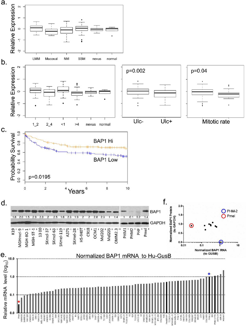
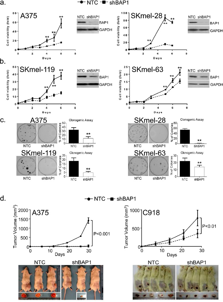
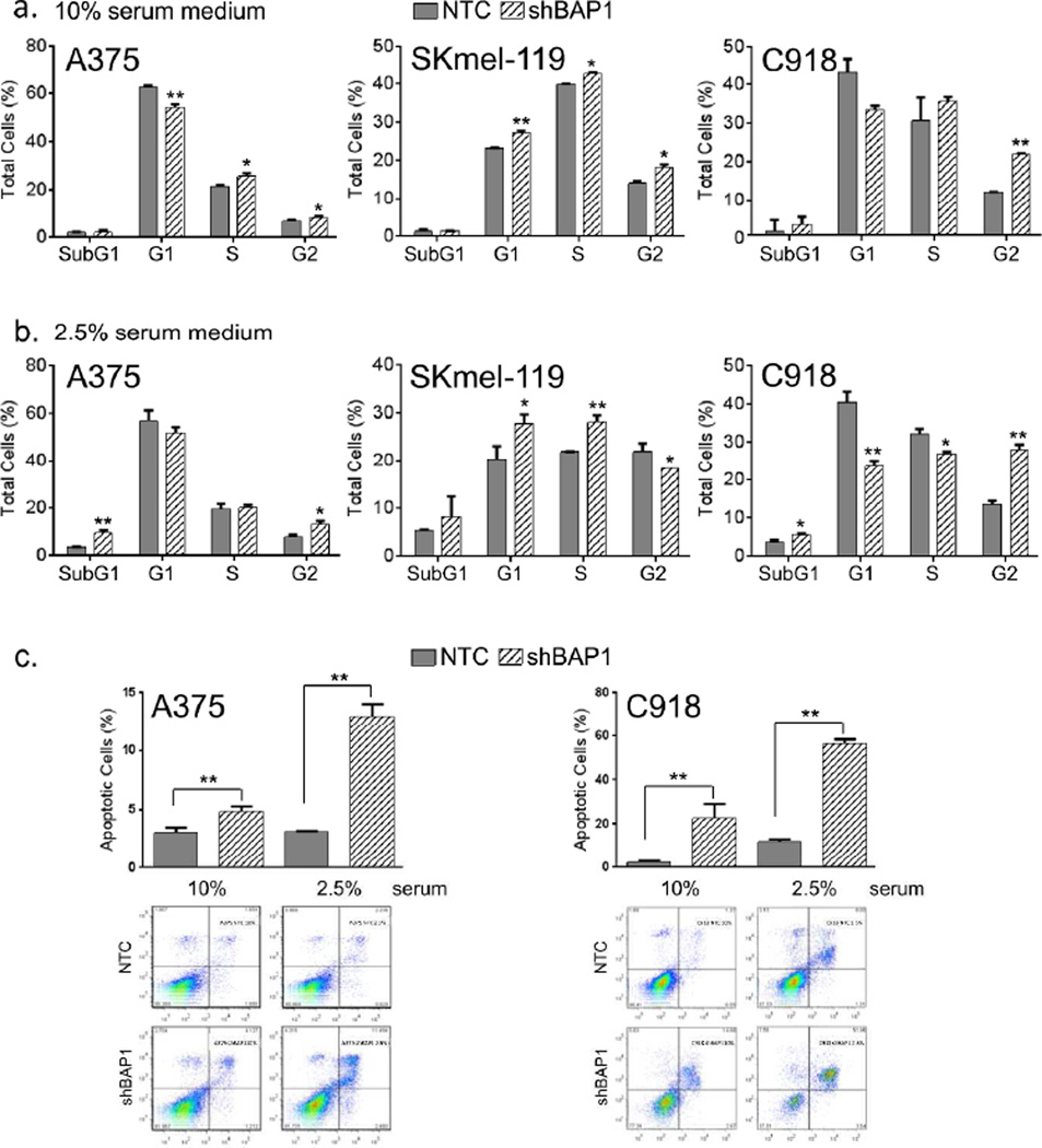
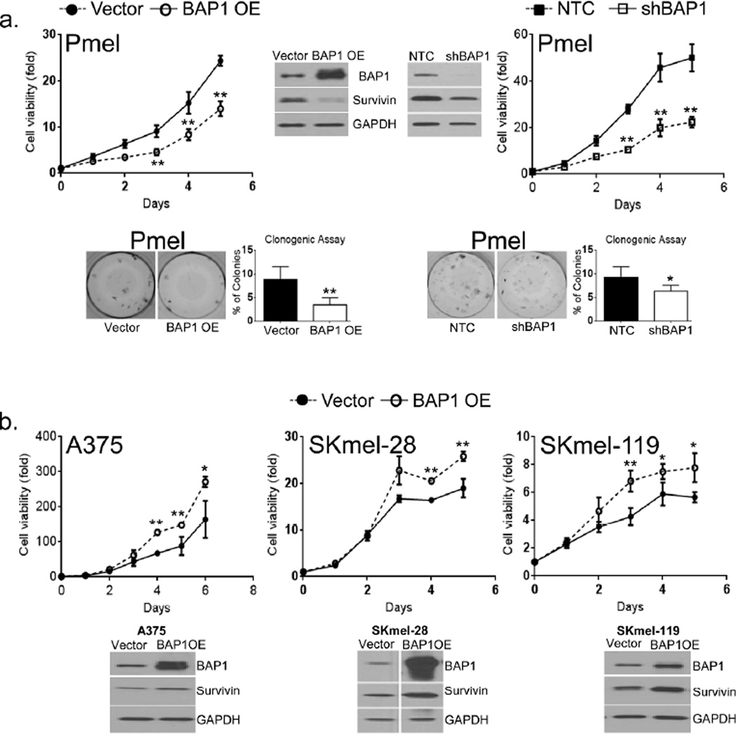
Although the pattern of BAP1 inactivation in ocular melanoma specimens and in the BAP1 cutaneous melanoma (CM)/ocular melanoma predisposition syndrome suggests a tumor suppressor function, the specific role of this gene in the pathogenesis of CM is not fully understood. We thus set out to characterize BAP1 in CM and discovered an unexpected pro-survival effect of this protein. Tissue and cell lines analysis showed that BAP1 expression was maintained, rather than lost, in primary melanomas compared with nevi and normal skin. Genetic depletion of BAP1 in melanoma cells reduced proliferation and colony-forming capability, induced apoptosis, and inhibited melanoma tumor growth in vivo. On the molecular level, suppression of BAP1 led to a concomitant drop in the protein levels of survivin, a member of anti-apoptotic proteins and a known mediator of melanoma survival. Restoration of survivin in melanoma cells partially rescued the growth-retarding effects of BAP1 loss. In contrast to melanoma cells, stable overexpression of BAP1 into immortalized but non-transformed melanocytes did suppress proliferation and reduce survivin. Taken together, these studies demonstrate that BAP1 may have a growth-sustaining role in melanoma cells, but that its impact on ubiquitination underpins a complex physiology, which is context and cell dependent.


Conflict of interest statement

Conflict of Interest

None of the authors have a conflict of interest related to the content of this manuscript.

Figures

Figure 1
Figure 1. BAP1 expression in primary cutaneous melanomas and melanoma lines
(a) BAP1 expression in histological types of melanoma. (b) BAP1 expression melanomas stratified by Breslow thickness, BAP1 expression in relation to ulceration and mitotic rate. (c) Survival differences between BAP1 high (>mean gene expression across the melanomas) and BAP1 low tumors (≤ mean gene expression across the melanomas). (d) Western blot analysis showing relative protein levels of BAP1 in a collection of 16 melanoma cell lines, primary human melanocytes (PHMs), primary human fibroblasts (PHF), and an immortalized non-transformed human melanocytes (Pmel). (e) Ranked BAP1 RNA levels (normalized to human GUSB) in a panel of melanoma lines and PHM-1, PHM-2 and Pmel. (f) Correlation between normalized BAP1 protein (to GAPDH) and normalized BAP1 RNA (to Hu-GUSB). Pmel (red circle and red asterisk) shows low RNA levels but strong protein expression while PHM-2 (blue circle and blue asterisk) shows high RNA levels with a near absence of detectable protein.
Figure 2
Figure 2. BAP1 depletion leads to melanoma growth suppression
The shRNA-mediated suppression of BAP1 results in reduced in vitro proliferation in 4 cutaneous melanoma cell lines. (a) A375 and SKmel-28 harbor BRAF (V600E) mutations while, (b) SKmel-119 and SKmel-63 contain NRAS (Q61R) and NRAS (Q61K) mutations, respectively. Error bars represent SEM from triplicate samples. (c) Loss of BAP1 is also associated with diminished colony forming capability. Error bars represent ±S.D., from at least 3 independent experiments. *p<0.05, **p<0.01 by student T test. (d) Effects of BAP1 depletion on tumor growth in a xenograft model. One million A375(NTC), A375(shBAP1), C918(NTC), and C918(shBAP1) cells were implanted subcutaneously in NOD-SCID-IL2G-null mice with matrigel in a 1:1 ratio and observed over the indicated time period. With A375 (cutaneous melanoma), 3 animals were used in each arm while for C918 (uveal melanoma), 5 animals were used in each arm. Error bars represent ± S.D., in tumor volume. *p<0.05, **p<0.01 by student T test.
Figure 3
Figure 3. BAP1 depletion causes cell cycle arrest and apoptosis
Analysis of cell cycle progression and apoptosis were performed at 6 or 7th day following shRNA-mediated BAP1 silencing. Cell cycle analysis in (a) 10% and (b) 2.5% serum medium using control cells (gray shading) and shBAP1 knockdown cells (hatched shading). Error bars represent SEM from triplicate samples and 3 experimental replicates are shown. A375 and SKmel-119 are cutaneous melanomas and C918 is an ocular melanoma used for comparison; *p<0.05, **p<0.01 by student T test. (c) FITC-Annexin staining of cultured control and BAP1-depleted cells in both 10% and 2.5% serum. DNA fragmentation may underestimate the level of apoptosis, especially if the total DNA content is elevated from G2/M arrest. Error bars represent SEM from triplicate samples both replicates shown; **p<0.01 by student T test.
Figure 4
Figure 4. BAP1 suppression is associated with survivin depletion
(a) RNA and protein levels of BIRC5 (survivin) upon BAP1 suppression in 4 melanoma lines. There is evidence of near total survivin loss at the protein level in A375, SKmel-28 and C918 but not SKmel-119. Error bars represent SEM from triplicate samples; *p<0.05 **p<0.01 by student T test. (b) Survivin over-expression (OE) in A375 cells led to the rescue of the in vitro growth arrest induced by shBAP1. (c) Schematic diagram of experimental design for ubiquitination assay. Indicated cell lines were incubated with MG132 (25 QM) for 6 hours, followed by removal of MG132 and the addition of cycloheximide (CHX, 25 Qg/ml) for the designated time intervals. “C” represents control cells that were exposed to neither MG132 nor CHX. “+M” indicates cells that were exposed to only MG132 and not CHX; for these cells, lysate was collected at time=0. “+M-C” represents cells that were exposed to MG132 and then switched to CHX; lysates were collected at 1, 2, 4 and 6 hrs post-CHX switch. “+M-D” represent cells that were exposed to MG132 and then DMSO control for 6 hours. (d) The effect of BAP1 depletion on survivin and GAPDH protein levels as measured by western blotting. If BAP1 directly deubiquitinated survivin, then survivin decay should be accelerated with BAP1 depletion. Even though the absolute levels of BAP1 appear to be lower in the shBAP1 lines, survivin degradation appears similar. Error bars represent SEM of triplicate samples. The experiments were performed 3 times with similar results; **p<0.01 by student T test.
Figure 5
Figure 5. BAP1 overexpression in Pmel and melanomas
(a) Effects of BAP1 overexpression (BAP1 OE) and depletion (shBAP1) on Pmel proliferation, survivin levels and colony forming capacity. Both BAP1 elevation and suppression in Pmel cells were associated with a decrease in colony formation, proliferation and levels of survivin. (b) Overexpression of BAP1 in 3 melanoma lines had more stimulatory effects on proliferation and survivin. The experiments were performed 3 times with similar results; *p<0.05, **p<0.01 by student T test.

References

    1. Abdel-Rahman MH, Pilarski R, Cebulla CM, et al. Germline BAP1 mutation predisposes to uveal melanoma, lung adenocarcinoma, meningioma, and other cancers. J Med Genet. 2011 - PMC - PubMed
    1. Bott M, Brevet M, Taylor BS, et al. The nuclear deubiquitinase BAP1 is commonly inactivated by somatic mutations and 3p21.1 losses in malignant pleural mesothelioma. Nat Genet. 2011 Jun 5;43(7):668–672. - PMC - PubMed
    1. Carbone M, Yang H, Pass HI, et al. BAP1 and cancer. Nature reviews Cancer. 2013;13:153–159. - PMC - PubMed
    1. Dalinghaus M, Rudolph CD, Rudolph AM. Effects of maternal fasting on hepatic gluconeogenesis and glucose metabolism in fetal lambs. Journal of developmental physiology. 1991;16:267–275. - PubMed
    1. Dey A, Seshasayee D, Noubade R, et al. Loss of the tumor suppressor BAP1 causes myeloid transformation. Science. 2012;337:1541–1546. - PMC - PubMed

Publication types

MeSH terms