

Normative amplitude-integrated EEG measures in preterm infants
- PMID: 25521561
- PMCID: PMC4447544
- DOI: 10.1038/jp.2014.225
Normative amplitude-integrated EEG measures in preterm infants
Abstract
Objective: Assessing qualitative patterns of amplitude-integrated electroencephalography (aEEG) maturation of preterm infants requires personnel with training in interpretation and an investment of time. Quantitative algorithms provide a method for rapidly and reproducibly assessing an aEEG recording independent of provider skill level. Although there are several qualitative and quantitative normative data sets in the literature, this study provides the broadest array of quantitative aEEG measures in a carefully selected and followed cohort of preterm infants with mild or no visible injury on term-equivalent magnetic resonance imaging (MRI) and subsequently normal neurodevelopment at 2 and 7 years of age.
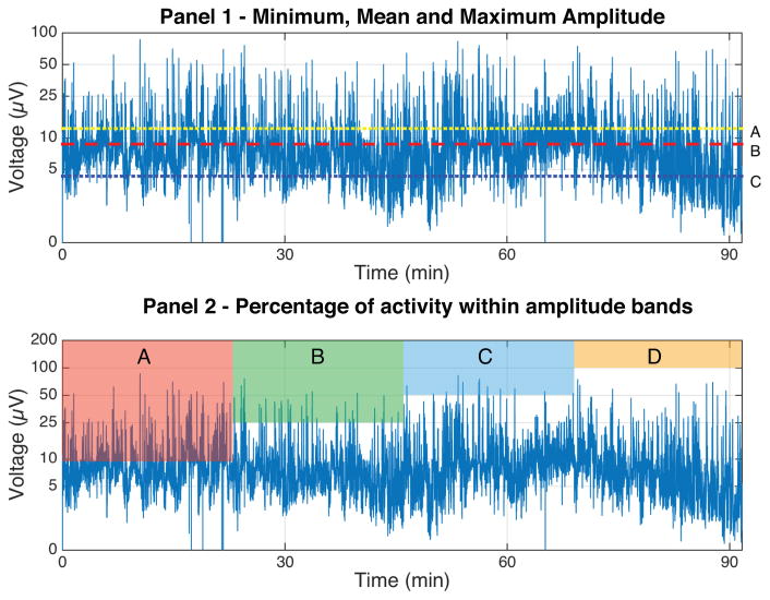
Study design: A two-channel aEEG recording was obtained on days 4, 7, 14 and 28 of life for infants born ⩽30 weeks estimated gestational age. Measures of amplitude and continuity, spectral edge frequency, percentage of trace in interburst interval (IBI), IBI length and frequency counts of smooth delta waves, delta brushes and theta bursts were obtained. MRI was obtained at term-equivalent age and neurodevelopmental testing was conducted at 2 and 7 years of corrected age.
Result: Correlations were found between increasing postmenstrual age (PMA) and decreasing maximum amplitude (R= -0.23, P=0.05), increasing minimum amplitude (R=0.46, P=0.002) and increasing spectral edge frequency (R=0.78, P=4.17 × 10(-14)). Negative correlations were noted between increasing PMA and counts of smooth delta waves (R= -0.39, P=0.001), delta brushes (R= -0.37, P=0.003) and theta bursts (R= -0.61, P=5.66 × 10(-8)). Increasing PMA was also associated with a decreased amount of time spent in the IBI (R= -0.38, P=0.001) and a shorter length of the maximum IBI (R= -0.27, P=0.03).
Conclusion: This analysis supports a strong correlation between quantitatively determined aEEG measures and PMA, in a cohort of preterm infants with normal term-equivalent age neuroimaging and neurodevelopmental outcomes at 7 years of age, which is both predictable and reproducible. These 'normative' quantitative values support the pattern of maturation previously identified by qualitative analysis.
Conflict of interest statement
The authors declare no conflicts of interest.
Figures
References
-
- Burdjalov VF, Baumgart S, Spitzer AR. Cerebral function monitoring: a new scoring system for the evaluation of brain maturation in neonates. Pediatrics. 2003 Oct;112(4):855–61. - PubMed
-
- Hellström-Westas L, De Vries LS, Rosén I. Atlas of amplitude-integrated EEGs in the newborn. London; Boca Raton, FL: Informa Healthcare_; Distributed in North and South America by Taylor & Francis; 2008.
-
- Vecchierini M-F, André M, d’ Allest AM. Normal EEG of premature infants born between 24 and 30 weeks gestational age: terminology, definitions and maturation aspects. Neurophysiol Clin Clin Neurophysiol. 2007 Nov;37(5):311–23. - PubMed
-
- West CR, Harding JE, Williams CE, Nolan M, Battin MR. Cot-side electroencephalography for outcome prediction in preterm infants: observational study. Arch Dis Child Fetal Neonatal Ed. 2011 Mar;96(2):F108–13. - PubMed
-
- Niemarkt HJ, Andriessen P, Peters CHL, Pasman JW, Blanco CE, Zimmermann LJ, et al. Quantitative analysis of amplitude-integrated electroencephalogram patterns in stable preterm infants, with normal neurological development at one year. Neonatology. 2010;97(2):175–82. - PubMed
Publication types
MeSH terms
Grants and funding
LinkOut - more resources
Full Text Sources
Other Literature Sources
Medical
