. 2014 Dec 16;4(4):1102-39.
doi: 10.3390/biom4041102.

Immunoglobulins: 25 years of immunoinformatics and IMGT-ONTOLOGY

Affiliations

Immunoglobulins: 25 years of immunoinformatics and IMGT-ONTOLOGY

Marie-Paule Lefranc. Biomolecules. .

Abstract

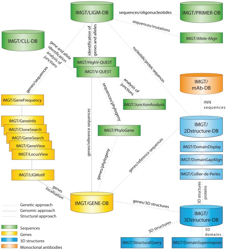
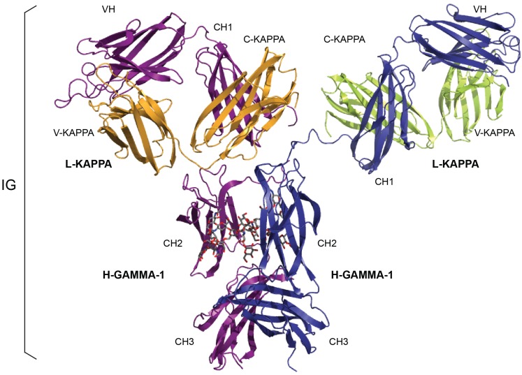
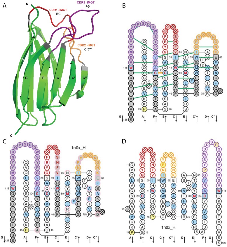
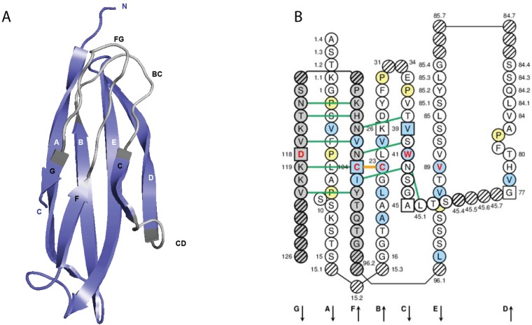
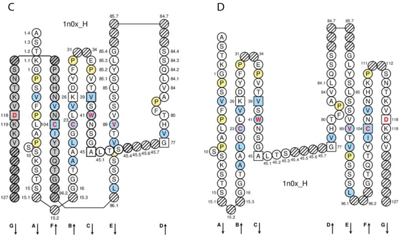
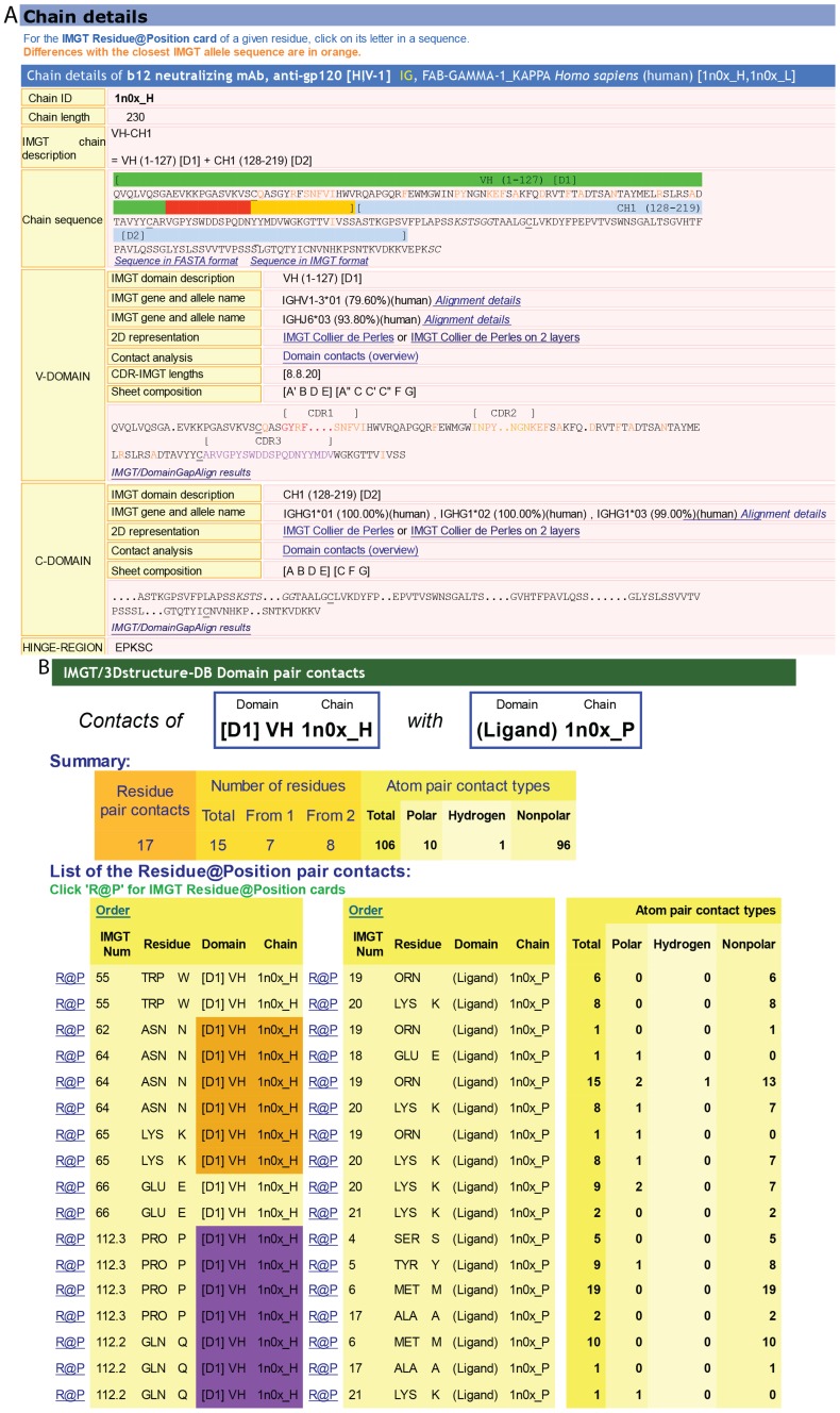
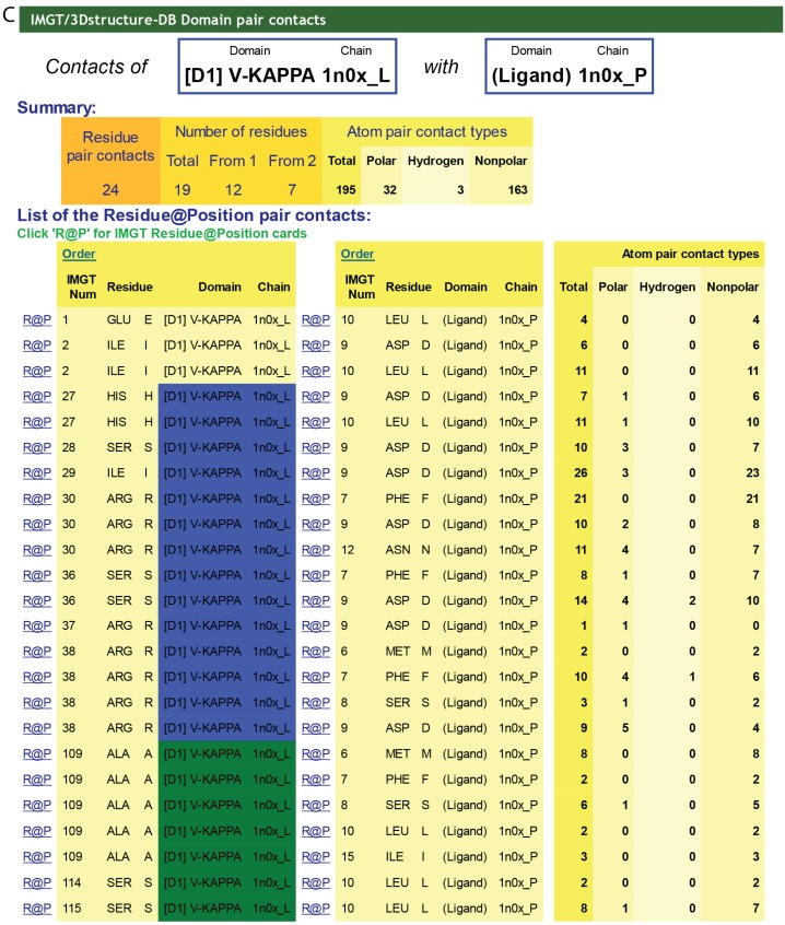
IMGT®, the international ImMunoGeneTics information system® (CNRS and Montpellier University) is the global reference in immunogenetics and immunoinformatics. By its creation in 1989, IMGT® marked the advent of immunoinformatics, which emerged at the interface between immunogenetics and bioinformatics. IMGT® is specialized in the immunoglobulins (IG) or antibodies, T cell receptors (TR), major histocompatibility (MH), and IgSF and MhSF superfamilies. IMGT® has been built on the IMGT-ONTOLOGY axioms and concepts, which bridged the gap between genes, sequences and three-dimensional (3D) structures. The concepts include the IMGT® standardized keywords (identification), IMGT® standardized labels (description), IMGT® standardized nomenclature (classification), IMGT unique numbering and IMGT Colliers de Perles (numerotation). IMGT® comprises seven databases, 15,000 pages of web resources and 17 tools. IMGT® tools and databases provide a high-quality analysis of the IG from fish to humans, for basic, veterinary and medical research, and for antibody engineering and humanization. They include, as examples: IMGT/V-QUEST and IMGT/JunctionAnalysis for nucleotide sequence analysis and their high-throughput version IMGT/HighV-QUEST for next generation sequencing, IMGT/DomainGapAlign for amino acid sequence analysis of IG domains, IMGT/3Dstructure-DB for 3D structures, contact analysis and paratope/epitope interactions of IG/antigen complexes, and the IMGT/mAb-DB interface for therapeutic antibodies and fusion proteins for immunological applications (FPIA).


Figures

Figure 1
Figure 1
IMGT®, the international ImMunoGeneTics information system® [1,2]. Databases are shown as cylinders and tools as rectangles. The web resources are not shown.
Figure 2
Figure 2
An immunoglobulin (IG) or antibody. In vivo, an IG or antibody is anchored in the membrane of a B cell as part of a signaling B cell receptor (BcR = membrane IG + CD79) or, as shown here, is secreted [3]. An IG is made of two identical heavy (H, for IG-HEAVY) chains and two identical light (L, for IG-LIGHT) chains [3]. An IG comprises 12 domains (for example, IgG1, shown here) or 14 domains (IgM or IgE). The V-DOMAIN of each chain (green online) and the C-DOMAIN, one for each L chain and three for each H chain (blue online) are highlighted. The light chain (here, L-KAPPA) is made of a variable domain (V-DOMAIN, here, V-KAPPA) at the N-terminal end and a constant domain (C-DOMAIN, here, C-KAPPA) at the C-terminal end. The heavy chain (here, H-GAMMA-1) is made of a VH (at the N-terminal end) and of three CH (four for H-MU or H-EPSILON) (Table 1) [3]. The structure is that of the antibody b12, an IgG1-kappa, and so far the only complete human IG crystallized (1hzh from IMGT/3Dstructure-DB [1]).
Figure 3
Figure 3
Variable (V) domain. An IG VH (V-DOMAIN) is shown as example. (A) 3D structure ribbon representation with the IMGT strand and loop delimitations [62]; (B) IMGT Collier de Perles on two layers with hydrogen bonds. The IMGT Collier de Perles on two layers show, in the forefront, the GFCC'C'' strands (forming the sheet located at the interface VH/VL of the IG) and, in the back, the ABED strands. The IMGT Collier de Perles with hydrogen bonds (green lines online, only shown here for the GFCC'C'' sheet) is generated by the IMGT/Collier-de-Perles tool integrated in IMGT/3Dstructure-DB, from experimental 3D structure data [9,10,11]; (C) IMGT Collier de Perles on two layers generated from IMGT/DomainGapAlign [10,25,26]. Pink circles (online) indicate amino acid changes compared to the closest genes and alleles from the IMGT reference directory; (D) IMGT Collier de Perles on one layer. Amino acids are shown in the one-letter abbreviation All proline (P) are shown online in yellow. IMGT anchors are in square. Hatched circles are IMGT gaps according to the IMGT unique numbering for V domain [62,65]. Positions with bold (online red) letters indicate the four conserved positions that are common to a V domain and to a C domain: 23 (1st-CYS), 41 (CONSERVED-TRP), 89 (hydrophobic), 104 2nd-CYS) [60,61,62,63,65], and the fifth conserved position, 118 (J-TRP or J-PHE) which is specific to a V-DOMAIN and belongs to the motif F/W-G-X-G that characterizes the J-REGION [62,65] (Table 2). The hydrophobic amino acids (hydropathy index with positive value: I, V, L, F, C, M, A) and tryptophan (W) [29] found at a given position in more than 50% of sequences are shown (online with a blue background color). Arrows indicate the direction of the beta strands and their designations in 3D structures. IMGT color menu for the CDR-IMGT of a V-DOMAIN indicates the type of rearrangement, V-D-J (for a VH, red, orange and purple) or V-J (for V-KAPPA or V-LAMBDA, blue, green and greenblue) [3]. The identifier of the chain to which the VH domain belongs is 1n0x_H (from the Homo sapiens b12 Fab) in IMGT/3Dstructure-DB [1]. The CDR-IMGT lengths of this VH are [8.8.20] and the FR-IMGT are [25.17.38.11]. The 3D ribbon representation was obtained using PyMOL [97] and “IMGT numbering comparison” of 1n0x_H (VH) from IMGT/3Dstructure-DB [1].
Figure 4
Figure 4
Constant (C) domain. An IG CH (C-DOMAIN) is shown as example. (A) 3D structure ribbon representation with the IMGT strand and loop delimitations [63]; (B) IMGT Collier de Perles on two layers with hydrogen bonds. The IMGT Colliers de Perles on two layers show, in the forefront, the GFC strands and, in the back, the ABED strands (located at the interface CH1/CL of the IG), linked by the CD transversal strand. The IMGT Collier de Perles with hydrogen bonds (green lines online, only shown here for the GFC sheet) is generated by the IMGT/Collier-de-Perles tool integrated in IMGT/3Dstructure-DB, from experimental 3D structure data [9,10,11]; (C) IMGT Collier de Perles on two layers from IMGT/DomainGapAlign [10,25,26]; (D) IMGT Colliers de Perles on one layer. Amino acids are shown in the one-letter abbreviation. All proline (P) are shown online in yellow. IMGT anchors are in square. Hatched circles are IMGT gaps according to the IMGT unique numbering for C domain [63,65]. Positions with bold (online red) letters indicate the four conserved positions that are common to a V domain and to a C domain: 23 (1st-CYS), 41 (CONSERVED-TRP), 89 (hydrophobic), 104 (2nd-CYS) [60,61,62,63,65] (Table 3), and position 118 which is only conserved in V-DOMAIN. The identifier of the chain to which the CH domain belongs is 1n0x_H (from the Homo sapiens b12 Fab, in IMGT/3Dstructure-DB, [1]). The 3D ribbon representation was obtained using PyMOL and “IMGT numbering comparison” of 1n0x_H (CH1) from IMGT/3Dstructure-DB [1].
Figure 4
Figure 4
Constant (C) domain. An IG CH (C-DOMAIN) is shown as example. (A) 3D structure ribbon representation with the IMGT strand and loop delimitations [63]; (B) IMGT Collier de Perles on two layers with hydrogen bonds. The IMGT Colliers de Perles on two layers show, in the forefront, the GFC strands and, in the back, the ABED strands (located at the interface CH1/CL of the IG), linked by the CD transversal strand. The IMGT Collier de Perles with hydrogen bonds (green lines online, only shown here for the GFC sheet) is generated by the IMGT/Collier-de-Perles tool integrated in IMGT/3Dstructure-DB, from experimental 3D structure data [9,10,11]; (C) IMGT Collier de Perles on two layers from IMGT/DomainGapAlign [10,25,26]; (D) IMGT Colliers de Perles on one layer. Amino acids are shown in the one-letter abbreviation. All proline (P) are shown online in yellow. IMGT anchors are in square. Hatched circles are IMGT gaps according to the IMGT unique numbering for C domain [63,65]. Positions with bold (online red) letters indicate the four conserved positions that are common to a V domain and to a C domain: 23 (1st-CYS), 41 (CONSERVED-TRP), 89 (hydrophobic), 104 (2nd-CYS) [60,61,62,63,65] (Table 3), and position 118 which is only conserved in V-DOMAIN. The identifier of the chain to which the CH domain belongs is 1n0x_H (from the Homo sapiens b12 Fab, in IMGT/3Dstructure-DB, [1]). The 3D ribbon representation was obtained using PyMOL and “IMGT numbering comparison” of 1n0x_H (CH1) from IMGT/3Dstructure-DB [1].
Figure 5
Figure 5
IMGT/3Dstructure-DB. (A) IMGT/3Dstructure-DB card. The “IMGT/3Dstructure-DB card” is available for each entry of the database. The “Chain details” shows, first, information on the chain (Chain ID, Chain length, IMGT chain description, Chain sequence), then a detailed description of each domain of the chain. The description of the V-DOMAIN (VH) and C-DOMAIN (CH1) of the VH-CH1 chain (1n0x_H) of the b12 Fab is shown. A similar result display interface is provided in IMGT/2Dstructure-DB cards but without “Contact analysis” (and without hydrogen bonds in IMGT Collier de Perles on 2 layers); (B) IMGT/3Dstructure-DB Domain pair contacts between the “VH” and the “Ligand” (antigen, Ag) of an IG/Ag complex. The VH is from the VH-CH1 chain (1n0x_H) of the b12 Fab and the ligand is a synthetic peptide (1n0x_P). The VH is in contact with the ligand by three AA of the CDR2-IMGT (orange online) (N62, N64 and K65) and two AA of the CDR3-IMGT (purple online) (P112.3 and Q112.2). The two AA which interact with the ligand but do not belong to the CDR-IMGT are the anchors W55 and E66. These contacts are not unexpected given by the small size (peptide) of the ligand; (C) IMGT/3Dstructure-DB Domain pair contacts between the “V-KAPPA” and the “Ligand” (Ag) of an IG/Ag complex. The V-KAPPA is from the L-KAPPA chain (1n0x_L) of the b12 Fab and the ligand is the peptide (1n0x_P) as in (B). The V-KAPPA is in contact with the ligand by seven AA of the CDR1-IMGT (blue online) (H27, S28, I29, R30, S36, R37 and R38) and three AA of the CDR3-IMGT (greenblue online) (A109, S114 and S115). “Polar”, “Hydrogen bond” and “Nonpolar” are selected by default in “Atom pair contact types” options at the bottom of the page (not shown). The user can also choose to display these contacts by “Atom pair contact categories” (BB), (SS), (BS) and (SB). Clicking on R@P gives access to the IMGT Residue@Position card. The IG/Ag complex structure is 1n0x from IMGT/3Dstructure-DB [1,9,10,11].
Figure 5
Figure 5
IMGT/3Dstructure-DB. (A) IMGT/3Dstructure-DB card. The “IMGT/3Dstructure-DB card” is available for each entry of the database. The “Chain details” shows, first, information on the chain (Chain ID, Chain length, IMGT chain description, Chain sequence), then a detailed description of each domain of the chain. The description of the V-DOMAIN (VH) and C-DOMAIN (CH1) of the VH-CH1 chain (1n0x_H) of the b12 Fab is shown. A similar result display interface is provided in IMGT/2Dstructure-DB cards but without “Contact analysis” (and without hydrogen bonds in IMGT Collier de Perles on 2 layers); (B) IMGT/3Dstructure-DB Domain pair contacts between the “VH” and the “Ligand” (antigen, Ag) of an IG/Ag complex. The VH is from the VH-CH1 chain (1n0x_H) of the b12 Fab and the ligand is a synthetic peptide (1n0x_P). The VH is in contact with the ligand by three AA of the CDR2-IMGT (orange online) (N62, N64 and K65) and two AA of the CDR3-IMGT (purple online) (P112.3 and Q112.2). The two AA which interact with the ligand but do not belong to the CDR-IMGT are the anchors W55 and E66. These contacts are not unexpected given by the small size (peptide) of the ligand; (C) IMGT/3Dstructure-DB Domain pair contacts between the “V-KAPPA” and the “Ligand” (Ag) of an IG/Ag complex. The V-KAPPA is from the L-KAPPA chain (1n0x_L) of the b12 Fab and the ligand is the peptide (1n0x_P) as in (B). The V-KAPPA is in contact with the ligand by seven AA of the CDR1-IMGT (blue online) (H27, S28, I29, R30, S36, R37 and R38) and three AA of the CDR3-IMGT (greenblue online) (A109, S114 and S115). “Polar”, “Hydrogen bond” and “Nonpolar” are selected by default in “Atom pair contact types” options at the bottom of the page (not shown). The user can also choose to display these contacts by “Atom pair contact categories” (BB), (SS), (BS) and (SB). Clicking on R@P gives access to the IMGT Residue@Position card. The IG/Ag complex structure is 1n0x from IMGT/3Dstructure-DB [1,9,10,11].

References

    1. IMGT®, the international ImMunoGeneTics information system®. [(accessed on 4 November 2014)]. Available online: http://www.imgt.org/
    1. Lefranc M.-P., Giudicelli V., Duroux P., Jabado-Michaloud J., Folch G., Aouinti S., Carillon E., Duvergey H., Houles A., Paysan-Lafosse T., et al. IMGT®, the international ImMunoGeneTics information system® 25 years on. Nucleic Acids Res. 2014 doi: 10.1093/nar/gku1056. - DOI - PMC - PubMed
    1. Lefranc M.-P., Lefranc G. The Immunoglobulin FactsBook. Academic Press; London, UK: 2001. pp. 1–458.
    1. Lefranc M.-P., Lefranc G. The T Cell Receptor FactsBook. Academic Press; London, UK: 2001. pp. 1–398.
    1. Lefranc M.-P. Nomenclature of the human immunoglobulin genes. In: Coligan J.E., Bierer B.E., Margulies D.E., Shevach E.M., Strober W., editors. Current Protocols in Immunology. John Wiley and Sons; Hoboken, NJ, USA: 2000. pp. 1–37.

Publication types

Substances