. 2015 Mar;77(3):416-24.
doi: 10.1038/pr.2014.207. Epub 2014 Dec 18.

Toll-like receptor-4 in human and mouse colonic epithelium is developmentally regulated: a possible role in necrotizing enterocolitis

Affiliations

Toll-like receptor-4 in human and mouse colonic epithelium is developmentally regulated: a possible role in necrotizing enterocolitis

Di Meng et al. Pediatr Res. 2015 Mar.

Abstract

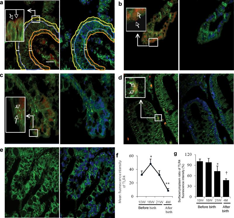
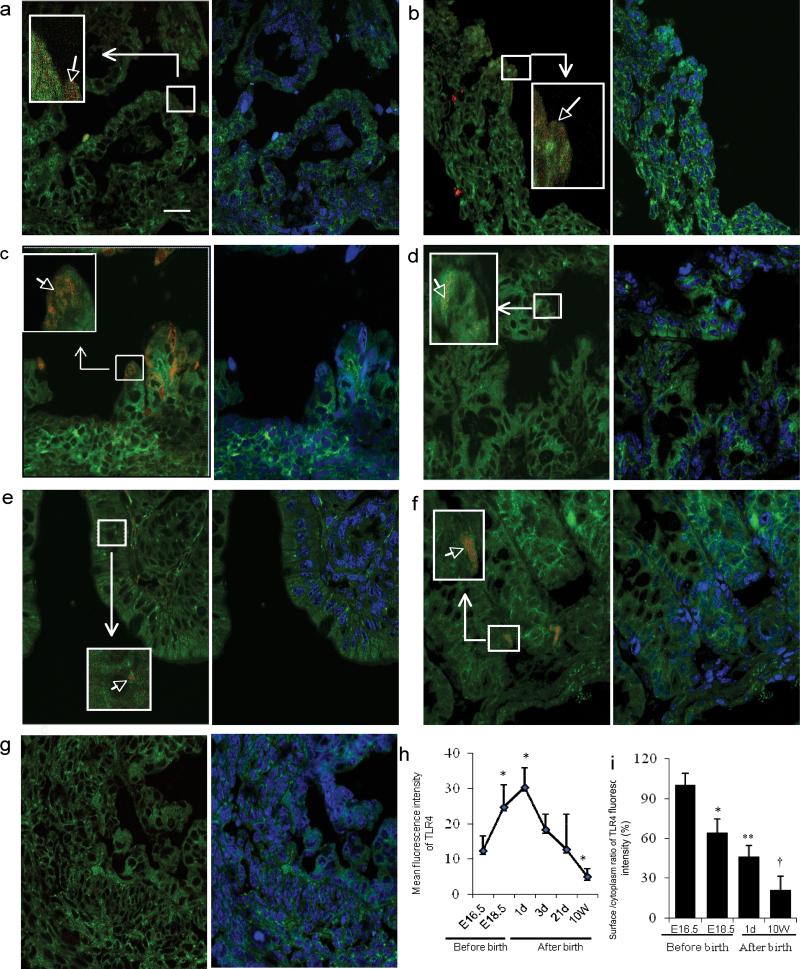
Background: Necrotizing enterocolitis (NEC) is an immature intestinal condition resulting in devastating intestinal inflammation due to unknown mechanisms. Evidence has suggested that intestinal maturation attenuates the severity of NEC and Toll-like receptor 4 (TLR4) has been suggested to play a critical role in its pathogenesis. We investigated whether maturational effects of TLR4 expression in immature colon might contribute to the development of NEC.

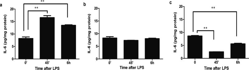
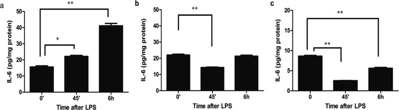
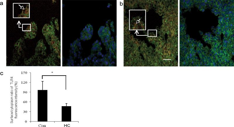
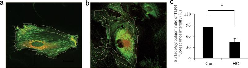
Methods: TLR4 colonocyte expression was detected by immunofluorescence confocal microscopy. Interleukin-6 (IL-6) levels were assayed by an enzyme-linked immunosorbent assay (ELISA).

Results: TLR4 expression was high in fetal colonic epithelium in human and mouse, with earlier gestation having a higher surface/cytoplasm distribution. TLR4 remained high in mouse postnatal day 1 but the surface/cytoplasm distribution was reduced. TLR4 decreased in amount and then was expressed in crypts in the mature human and mouse colon. Hydrocortisone (HC) reduced the surface/cytoplasm distribution of TLR4 in human fetal colon. Elevated IL-6 levels in immature colon after lipopolysaccharide were attenuated by HC in human and mouse.

Conclusion: Expression, localization, and signaling of TLR4 in colonic epithelium may be developmentally regulated. HC may accelerate the TLR developmental pathway change to an adult type, which may account for its impact on TLR4 signaling.


Figures

Figure 1
Figure 1
Location and quantity of TLR4 expression in human colonic epithelium from 10 weeks gestation to 4 months postpartum. Confocal microscopy image (a-e) left pane: overlayer of human colon tissues stained with a phycoerythrin (PE) labeled anti-human TLR4 antibody (red) and a fluorescein isothiocyanate (FITC) labeled anti- phospholipid antibody (green) (as a cell membrane marker); right pane: overlayer of phospholipid-FITC and DRAQ5(blue)(as a cell nuclear marker). Magnification × 1000; scale bar-20μm. (a) An area classification of colonic epithelium in a 10 week human colon. The thickness of the surface (line 1) and basal membrane (line 2) both are 3.75 μm. Surface area is surrounded by a yellow line, cytoplasm area is surrounded by a blue line and basal area was surrounded by a dark yellow line. (b) TLR4 expression at gestational age 16 weeks, (c) 21 weeks and (d) 4 months after birth. (e) Negative control for anti-human TLR4 antibody in a human fetal 16 week colonic section. (f) The average mean fluorescence intensity of TLR4 expression within the whole epithelial area (n=3-4) for each group, error bars represent SD, compared to a 10-week gestational age group * p<0.05, **p<0.01). (g) Histogram of surface to cytoplasm ratio of TLR4 fluorescence intensity in colonic sections (n=3-4) for each group, error bars represent SD, compare to a 10-week gestational age group * p<0.05, †P<0.001).
Figure 2
Figure 2
The ontogeny of TLR4 expression in mouse colonic epithelium during gestation and the postpartum period. Representative confocal image (a-g) left pane: overlayer of mouse colonic tissue stained with a PE labeled anti-mouse TLR4 antibody (red) and counter stained with FITC labeled anti-phospholipids antibody as a cell membrane marker (green). right pane: overlayer of phospholipid-FITC and DRAQ5(blue)(as a cell nuclear marker). The colonic epithelium classification uses the same measurement units as shown in Figure 1-a, but with 1.75 μm as the thickness of surface and basal membranes. Magnification × 1000; scale bar-20μm. (a) Representative image of TLR4 expression E16.5, (b) E18.5 and, (c) postnatal days 1, (d) 3, (e) 21 and 10 weeks (f). (g) Negative control of an anti-mouse TLR4 in an E18.5 mouse fetal colonic section. (h) The average mean fluorescent intensity of TLR4 expression within the entire epithelial area (n=3-4 for each group, error bars represent SD, * p<0.05 vs. E16.5). (i) Histogram of the surface to cytoplasm ratio of TLR4 fluorescent intensity in mouse colonic sections (n=3-4 for each group, error bars represent SD, * p<0.05, **p<0.01, †P<0.001 vs. E16.5).
Figure 3
Figure 3
Hydrocortisone (HC) induces a shift in the TLR4 response to LPS in human fetal colonic epithelial cells. FHC with or without HC pretreatment and adult human colonic epithelial cells (NCM 460) were stimulated with LPS for 45 minutes and for 6 hours (a) a FHC cell line, (b) FHC pre-treated with HC (1μM) for 5 days, and (c) a NCM 460 cell line. The means and SD are from triplicate wells and are representative of three separate experiments, **p<0.01 vs. 0 minute.
Figure 4
Figure 4
Hydrocortisone (HC) induced a shift in the response of TLR4 to LPS in mouse fetal colonic organ culture. C57BL/6J fetal and adult mouse colonic tissues were incubated with LPS for 45 minutes and for 6 hours and IL-6 levels measured in the supernatant (a) E 18.5, (b) E18.5 from a mother treated with HC, and (c) 12 week adult mouse colonic tissues. The mean and SD are from triplicate wells, and are representative of three separate experiments, *p<0.05, **p<0.01vs.0 minute.
Figure 5
Figure 5
Hydrocortisone (HC) reduces surface TLR4 expression in human fetal colonic epithelium. Human 16 week fetal colonic tissues incubated for 2 hours and then stimulated with HC or not for 20 hours. TLR4 expression was detected by the methods described. Original magnification ×1000; scale bar-20 μm. (a) TLR4 expression in control colonic sections. (b) HC pre-treated colonic sections. (c) Histogram of the surface to cytoplasm ratio of TLR4 fluorescent intensity in control and HC pre-treated colonic sections. The results represent the mean±SD, n=3, *p<0.05 vs. control group.
Figure 6
Figure 6
Hydrocortisone (HC) reduces surface to cytoplasm ratio of TLR4 in fetal human colonic epithelial cells. Original magnification ×1000; scale bar-20 μm measured as previously described. (a) Control FHC cells. (b) HC pre-treated FHC cells. (c) Histogram of the surface to cytoplasm ratio of TLR4 fluorescence intensity in control and HC pre-treated FHC cells, results represent a mean±SD, n=10, †p<0.001 vs. control group.

References

    1. Neu J, Walker WA. Necrotizing enterocolitis. N Engl J Med. 2011;364:255–264. - PMC - PubMed
    1. Grave GD, Nelson SA, Walker WA, et al. New therapies and preventive approaches for necrotizing enterocolitis: report of a research planning workshop. Pediatr Res. 2007;62:510–514. - PubMed
    1. Afrazi A, Sodhi CP, Hackam DJ. New insights into the pathogenesis and treatment of necrotizing enterocolitis: toll-like receptors and beyond. Pediatr Res. 2011;69:183–188. - PMC - PubMed
    1. Kosloske AM. Epidemiology of necrotising enterocolitis. Acta Paediatr Suppl. 1994;396:2–7. - PMC - PubMed
    1. Beeby PJ, Jeffery H. Risk factors for necrotising enterocolitis: the influence of gestational age. Arch Dis Child. 1992;67:432–435. - PMC - PubMed

Publication types

MeSH terms

LinkOut - more resources