Review
. 2014 Dec 16;15(12):23377-88.
doi: 10.3390/ijms151223377.

RNA recognition and stress granule formation by TIA proteins

Affiliations
Review

RNA recognition and stress granule formation by TIA proteins

Saboora Waris et al. Int J Mol Sci. .

Abstract

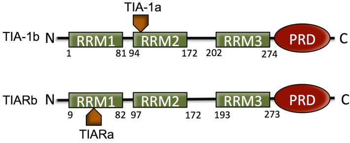
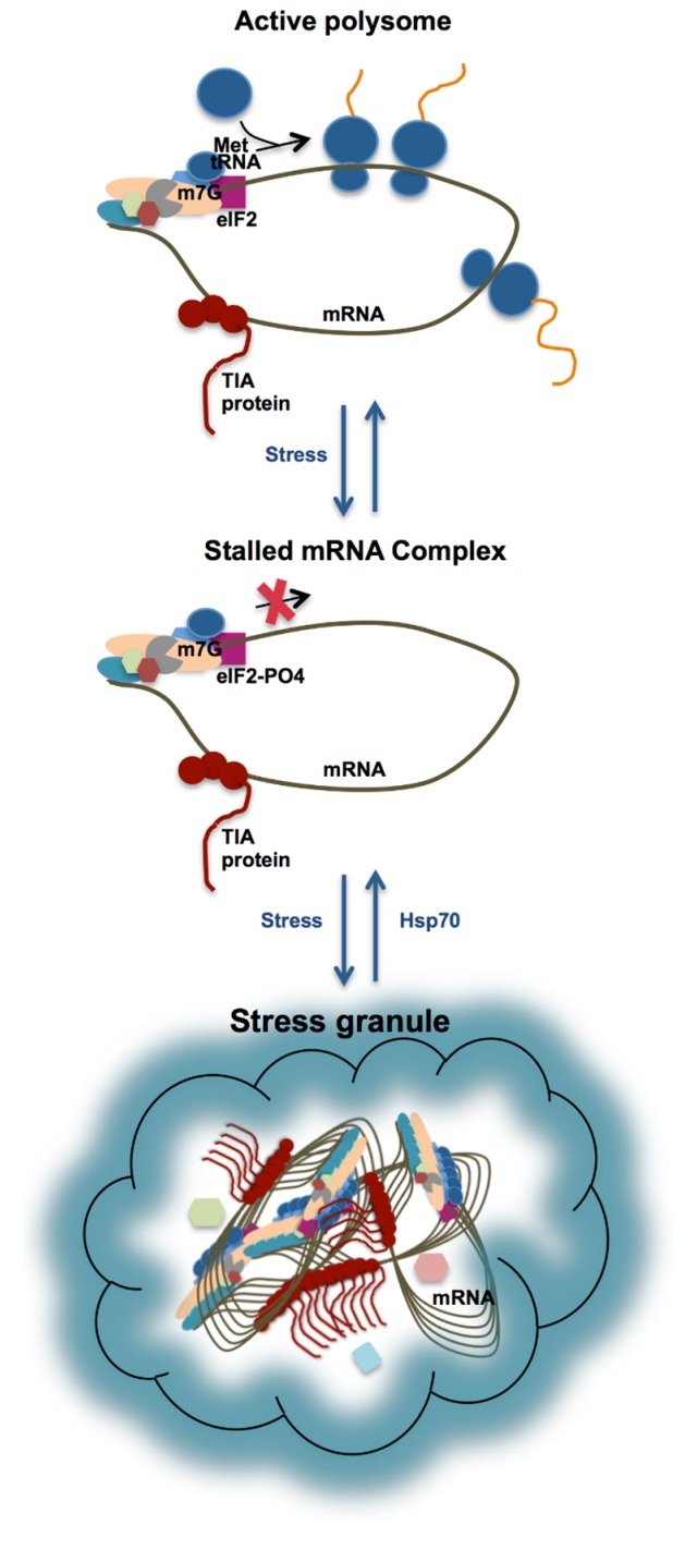
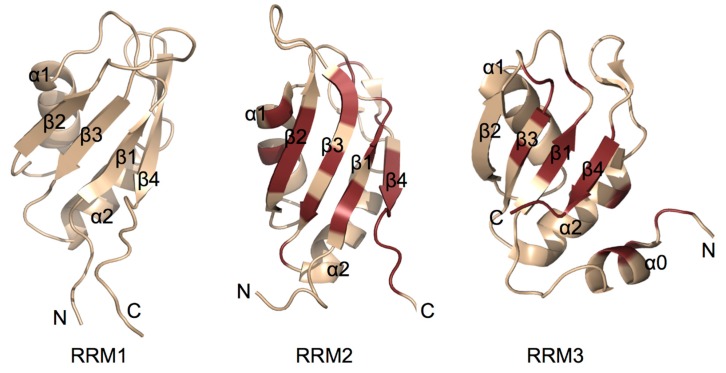
Stress granule (SG) formation is a primary mechanism through which gene expression is rapidly modulated when the eukaryotic cell undergoes cellular stresses (including heat, oxidative, viral infection, starvation). In particular, the sequestration of specifically targeted translationally stalled mRNAs into SGs limits the expression of a subset of genes, but allows the expression of heatshock proteins that have a protective effect in the cell. The importance of SGs is seen in several disease states in which SG function is disrupted. Fundamental to SG formation are the T cell restricted intracellular antigen (TIA) proteins (TIA-1 and TIA-1 related protein (TIAR)), that both directly bind to target RNA and self-associate to seed the formation of SGs. Here a summary is provided of the current understanding of the way in which TIA proteins target specific mRNA, and how TIA self-association is triggered under conditions of cellular stress.


Figures

Figure 1
Figure 1
Schematic of the domain structure of T cell restricted intracellular antigen (TIA) protein isoforms. TIA proteins consist of three RNA recognition motifs (RRMs) that provide RNA/DNA binding specificity and a Q-rich prion-related domain (PRD) domain, involved in stress granule (SG) formation. The two isoforms of both proteins that form as a result of alternative splicing are indicated.
Figure 2
Figure 2
Schematic of SG formation formed via the self-association of TIA proteins (shown in brown with circles representing RRM domains, and a tail representing the C-terminal PRD) bound to stalled ribonucleoprotein-mRNA complexes. Upon phosphorylation of eIF2 under conditions of stress, the pre-initiation complex is stalled. Accumulation of the stalled mRNA complex results in its incorporation into SG (involving interactions between the TIA protein PRDs) until such time that heat shock proteins are able to reverse the process and translation is resumed.
Figure 3
Figure 3
Cartoon representation of TIA protein RNA-recognition motif (RRM) structures solved using NMR spectroscopy. RRM 1 from TIAR (PDB entry:2CQI) and RRMs 2 and 3 from TIA-1 (PDB entry:2MJN) are shown. Amino acid residues of RRMs 2 and 3 shown to be perturbed upon binding by ssRNA are highlighted in ruby showing that binding mainly occurs across the surface the β-sheet, but also involves residues outside the canonical RRM fold [42,44,46,49]. No evidence for specific binding of RNA by RRM1 has been observed.

References

    1. Buchan J.R., Parker R. Eukaryotic stress granules: The ins and outs of translation. Mol. Cell. 2009;36:932–941. - PMC - PubMed
    1. Anderson P., Kedersha N. Stress granules. Curr. Biol. 2009;19:R397–R398. - PubMed
    1. Scheu S., Stetson D.B., Reinhardt R.L., Leber J.H., Mohrs M., Locksley R.M. Activation of the integrated stress response during T helper cell differentiation. Nat. Immunol. 2006;7:644–651. - PubMed
    1. Kim W.J., Back S.H., Kim V., Ryu I., Jang S.K. Sequestration of TRAF2 into stress granules interrupts tumor necrosis factor signaling under stress conditions. Mol. Cell. Biol. 2005;25:2450–2462. - PMC - PubMed
    1. Arimoto K., Fukuda H., Imajoh-Ohmi S., Saito H., Takekawa M. Formation of stress granules inhibits apoptosis by suppressing stress-responsive MAPK pathways. Nat. Cell Biol. 2008;10:1324–1332. - PubMed

Publication types