. 2014 Dec 18;10(12):e1004768.
doi: 10.1371/journal.pgen.1004768. eCollection 2014 Dec.

SCL, LMO1 and Notch1 reprogram thymocytes into self-renewing cells

Affiliations

SCL, LMO1 and Notch1 reprogram thymocytes into self-renewing cells

Bastien Gerby et al. PLoS Genet. .

Abstract

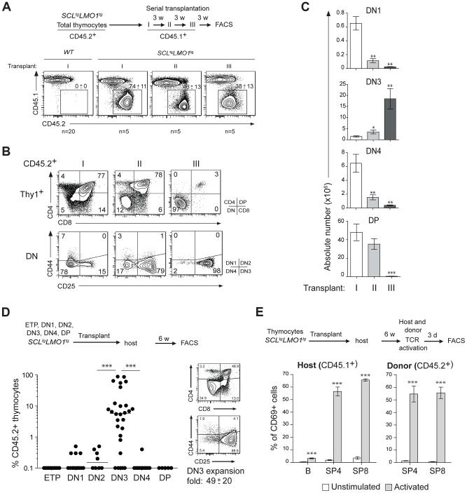
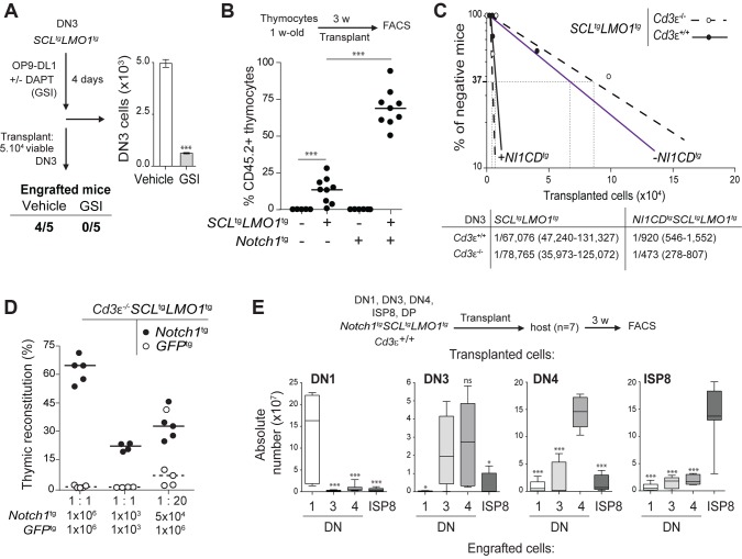
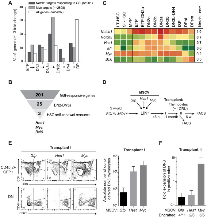
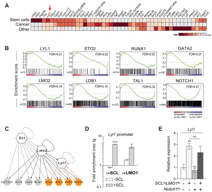
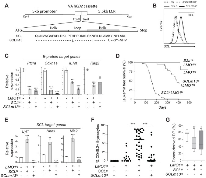
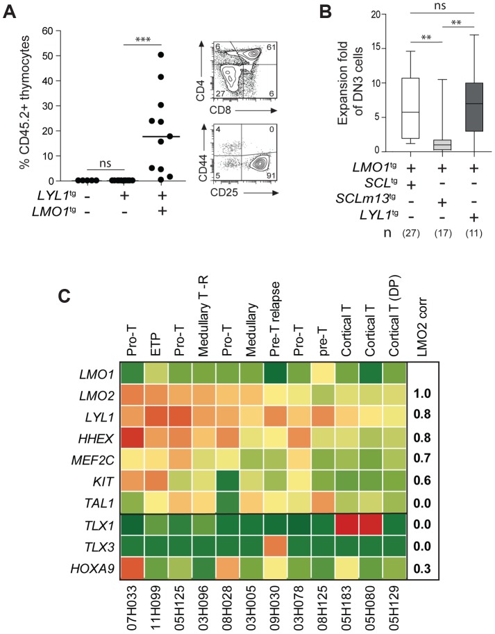
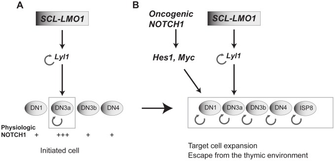
The molecular determinants that render specific populations of normal cells susceptible to oncogenic reprogramming into self-renewing cancer stem cells are poorly understood. Here, we exploit T-cell acute lymphoblastic leukemia (T-ALL) as a model to define the critical initiating events in this disease. First, thymocytes that are reprogrammed by the SCL and LMO1 oncogenic transcription factors into self-renewing pre-leukemic stem cells (pre-LSCs) remain non-malignant, as evidenced by their capacities to generate functional T cells. Second, we provide strong genetic evidence that SCL directly interacts with LMO1 to activate the transcription of a self-renewal program coordinated by LYL1. Moreover, LYL1 can substitute for SCL to reprogram thymocytes in concert with LMO1. In contrast, inhibition of E2A was not sufficient to substitute for SCL, indicating that thymocyte reprogramming requires transcription activation by SCL-LMO1. Third, only a specific subset of normal thymic cells, known as DN3 thymocytes, is susceptible to reprogramming. This is because physiological NOTCH1 signals are highest in DN3 cells compared to other thymocyte subsets. Consistent with this, overexpression of a ligand-independent hyperactive NOTCH1 allele in all immature thymocytes is sufficient to sensitize them to SCL-LMO1, thereby increasing the pool of self-renewing cells. Surprisingly, hyperactive NOTCH1 cannot reprogram thymocytes on its own, despite the fact that NOTCH1 is activated by gain of function mutations in more than 55% of T-ALL cases. Rather, elevating NOTCH1 triggers a parallel pathway involving Hes1 and Myc that dramatically enhances the activity of SCL-LMO1 We conclude that the acquisition of self-renewal and the genesis of pre-LSCs from thymocytes with a finite lifespan represent a critical first event in T-ALL. Finally, LYL1 and LMO1 or LMO2 are co-expressed in most human T-ALL samples, except the cortical T subtype. We therefore anticipate that the self-renewal network described here may be relevant to a majority of human T-ALL.


Conflict of interest statement

The authors declare no competing financial interest.

Figures

Figure 1
Figure 1. The SCL and LMO1 oncogenes confer an aberrant self-renewal potential to DN3 pre-leukemic thymocytes.
(A–C) Pre-leukemic SCL tg LMO1 tg thymocytes exhibit an aberrant self-renewal potential. Pre-leukemic SCL tg LMO1 tg thymocytes (CD45.2+) were serially transplanted into primary (I), secondary (II) and tertiary (III) recipient mice (CD45.1+) (1.5×10⁷ cells/mouse, 5 mice per group). Donor-derived thymocytes (CD45.1-CD45.2+) in the thymus were analyzed by flow cytometry 3 weeks after each transplantation. Note the absence of engrafment of wild type (WT) thymocytes when transplanted into primary mice (A). Immunophenotype of donor-derived thymocytes was assessed by flow cytometry (FACS) (B) and the absolute numbers of donor-derived DN1, DN3, DN4 and DP thymocytes were calculated after each transplantation (C). (D) SCL-LMO1-induced self-renewal activity is almost exclusively present in DN3 thymocytes. Purified thymocyte subpopulations (ETP, DN1-4, DP) from SCL tg LMO1 tg mice were transplanted (5×10⁴ cells per mouse). Recipient mice were analyzed for engraftment as above (left panel). Representative flow cytometry profiles of thymocytes generated by transplanted DN3 cells (right panel). There was a net 49-fold amplification of DN3 cells in vivo. (E) Engrafted SCL tg LMO1 tg thymocytes generate functional T cells in vivo that respond to TCR activation. Purified T cells were stimulated (activated) or not (control) with anti-CD3/CD28 beads and analyzed within the donor-derived SP4 and SP8 cells for expression of the CD69 activation marker. Host T and B cells served as positive and negative controls, respectively.
Figure 2
Figure 2. Notch1 collaborates with SCL-LMO1 to increase the pool of pre-LSCs and their competitiveness independently of a functional pre-TCR.
(A) The engraftment of SCL-LMO1 DN3 thymocytes is abrogated by γ-secretase inhibitor (GSI) treatment prior to transplantation. DN3 thymocytes were purified from pre-leukemic SCL tg LMO1 tg mice and co-cultured on OP9-DL1 stromal cells in the presence or absence (vehicle) of 2.5 µM DAPT (GSI) for 4 days. The total numbers of viable cells recovered per culture are shown (right panel). Following drug treatment, equal numbers of viable cells were transplanted (5×10⁴ per mouse, n = 5). Engrafted mice: number of positive mice showing thymic reconstitution per group. (B) A hyperactive Notch1 allele is insufficient to induce aberrant self-renewal in thymocytes but significantly enhances the engraftment of SCL tg LMO1 tg thymocytes. Total thymocytes (1.5×10⁷) from 1-week-old mice of the indicated genotype were transplanted; recipient mice were analyzed for thymic engraftment 3 weeks later. (C) Oncogenic Notch1 increases the frequencies of SCL-LMO1 pre-LSCs independently of a functional pre-TCR. Purified DN3 thymocytes from SCL tg LMO1 tg and Notch1 tg SCL tg LMO1 tg mice with (Cd3ε +/+) or without (Cd3ε -/-) a functional pre-TCR were transplanted in limiting dilution assays (upper panel). Mice were scored positive when T-cell lineage reconstitution was more than 1%; pre-LSC frequencies and confidence intervals (lower panel) were calculated by applying Poisson statistics using the Limiting Dilution Analysis software (StemCell Technologies). (D) Cd3ε -/- Notch1 tg SCL tg LMO1 tg pre-leukemic thymocytes outcompete Cd3ε -/- SCL tg LMO1 tg thymocytes. Reconstitution by Cd3ε -/- Notch1 tg SCL tg LMO1 tg (CD45.2+ GFP-, closed circles) and GFP tg Cd3ε -/- SCL tg LMO1 tg (CD45.2+ GFP+, open circles) thymocytes transplanted with the indicated cell numbers at 1∶1 or 1∶20 ratio. (E) Notch1 expands the cellular targets of SCL-LMO1 to DN1-4 and ISP8 cells. Pre-leukemic thymocyte subsets (DN1-4, ISP8 and DP) were purified from Notch1tgSCLtgLMO1 tg mice and transplanted at 5×10⁴ cells per recipient mouse. The absolute numbers of donor-derived DN1-4 and ISP8 cells was calculated for each transplantation.
Figure 3
Figure 3. Functional importance of Hes1 and c-Myc downstream of Notch1 in thymocyte reprogramming induced by SCL-LMO1.
(A) Expression of GSI-responsive NOTCH1 target genes during thymocyte differentiation. Global gene expression data of thymocyte subpopulations were obtained from the Immunological Genome Project (http://www.immgen.org/). The percentage of GSI-responsive NOTCH1 target genes or MYC target genes that are up-regulated at each transitional stage during thymocyte differentiation (>1.3-fold change) are shown. (B) Filtering of GSI-responsive NOTCH1 target genes that increase at the DN2 to DN3a transition and are present in HSC self-renewal resources (www.bioinfo.iric.ca/self-renewal/Main and www.bonemarrowhsc.com). (C) Gene expression profiles of Notch1 and their target genes (Notch3, Hes1, IL7r, Myc and Bcl6) during thymocyte differentiation were collected from the Immunological Genome Project and represented as a heat map. (D) Schematic strategy to study the role of Hes1 and c-Myc in self-renewal activity induced by SCL-LMO1. Lineage negative (LIN-) cells from SCL tg LMO1 tg mice (CD45.2+) were transduced with either MSCV-Hes1 and MSCV- Myc retroviral vectors or with control MSCV-GFP. Equal number (5×10⁴ cells) of purified GFP+LIN- cells were then transplanted in primary mice (CD45.2-). Donor-derived GFP+CD45.2+ thymocytes were transplanted at the limiting dose of ∼1 CRU (105 cells) per mouse into secondary recipients. (E) Immunophenotype of donor-derived GFP+CD45.2+ thymocytes in primary mice was analyzed by FACS (left panel) and the absolute number of DN3 cells was calculated (right panel). (F) The fold expansion of donor-derived GFP+CD45.2+ DN3 thymocytes was calculated in secondary mice.
Figure 4
Figure 4. Lyl1 coordinates a self-renewal network downstream of SCL-LMO1.
(A) Analysis of SCL-LMO1-upregulated genes in Cd3ε -/- thymocytes. Gene signatures were analysed using the Stem Cell Discovery Engine tool as described in Experimental procedures, and signatures deemed enriched in SCL-LMO1 up-regulated genes (adjusted p-val <0.05) were classified into broad categories. The heatmap depicts the frequency of association to each gene by signature categories (stem cells, cancer, and other). (B) GSEA analysis of hematopoietic transcription factor signatures in SCL-LMO1 thymocytes. The lists of genes bound by 31 hematopoietic transcription factors within 2 kb of their proximal promoters were extracted from a compendium of ChIP-seq experiments (see Materials and Methods). The top 7 transcription factors are illustrated (FDR, false discovery rate, ranging from 0.01–0.32). In comparison, NOTCH1-bound genes were not up- or down-regulated by SCL-LMO1. (C) Hierarchical organisation of the self-renewal network controlled by SCL-LMO1. Integration of published ChIP-seq data with up-regulated genes in DN3 pre-leukemic thymocytes identified common targets of SCL, LMO2 and LYL1 (highlighted in yellow). Incoming edges represent the binding of regulators at the proximal promoters of target genes (peaks within 2kb of the transcription initiation sites). (D) SCL and LMO1 occupy Lyl1 regulatory sequences. Chromatin extracts from the AD10.1 DN cell line expressing SCL (+SCL) or not (-SCL) were immunoprecipitated with the indicated antibodies. Lyl1 regulatory sequences were amplified by q-PCR. Data are expressed as fold enrichment over IgG controls. (E) Lyl1 gene expression is induced by SCL-LMO1 but is not modified by Notch1 in DN3 thymocytes. mRNA levels in purified DN3 thymocytes from the indicated transgenic mice were determined by qRT-PCR and normalized to β-Actin (Mean +/- SD, n = 3).
Figure 5
Figure 5. Transcription activation driven by SCL-LMO1 interaction is critical for thymocyte reprogramming and T-ALL induction.
(A) Generation of transgenic mice expressing the LMO1-binding defective mutant SCLm13. The sequence coding for wild type human SCL or human SCLm13 HLH domain mutant were cloned into the VA hCD2 cassette to generate transgenic mice. Shown are amino acids of the HLH region of SCL or SCLm13. (B) Immunofluorescence of human SCL (wt or m13) by flow cytometry. Thymocytes were stained with the monoclonal antibody against human SCL (BTL73). Control cells were stained with the second antibody only. (C) Expression of E protein target genes is inhibited both by SCL-LMO1 and SCLm13-LMO1 transgenes in DN3 thymocytes. mRNA levels in purified DN3 thymocytes from the indicated transgenic mice were determined by qRT-PCR and normalized to β-Actin (Mean +/- SD, n = 3). (D) Kaplan-Meier curves of the time to leukemia for LMO1tg, E2a+/-LMO1tg, SCLtgLMO1tg and SCLm13tgLMO1tg mice. (E) The interaction between SCL and LMO1 is required to activate the transcription of the self-renewal genes Lyl1, Hhex and Nfe2 in DN3 thymocytes. mRNA levels in purified DN3 thymocytes from the indicated transgenic mice were determined by qRT-PCR and normalized to β-Actin (Mean +/- SD, n = 3). (F–G) SCL but not the LMO1-binding defective SCL-m13 mutant collaborates with LMO1 to induce abnormal thymic reconstitution potential to thymocytes. Pre-leukemic thymocytes (1.5×10⁷ cells) from 3-week-old mice were transplanted. Recipient mice were analysed for thymic reconstitution (CD45.2+Thy1+) after 6 weeks (F) and the proportion of DP cells in engrafted CD45.2+Thy1+ thymocytes was assessed by FACS (G).
Figure 6
Figure 6. LYL1 and LMO1/2 are co-expressed in human T-ALL and collaborate to reprogram thymocytes.
(A) LYL1 collaborates with LMO1 to induce abnormal thymic reconstitution potential to thymocytes. 1.5×10⁷ thymocytes from the indicated mice were transplanted and thymic engraftment was analyzed after 6 weeks (left panel). Representative FACS profile of engrafted LYL1 tg LMO1 tg thymocytes (right panel). (B) LYL1-LMO1-induced DN3 expansion was comparable to SCL-LMO1-induced expansion after transplantation, as illustrated by the box plots (with the median and extreme values of each distribution, cohorts of n mice). (C) LMO2 expression levels correlate with LYL1 levels in T-ALL patient samples. Illustrated are the RPKM values for the indicated human gene. Note the high correlation coefficient between LMO2 and LYL1 and the absence of correlation with TLX1/3.
Figure 7
Figure 7. Model of the collaboration between the SCL, LMO1 and Notch1 oncogenes.
(A) SCL and LMO1 interact to upregulate Lyl1 gene expression and create a feed forward loop that activates self-renewal in DN3 thymocytes. DN3 cells are prone to SCL-LMO1 self-renewal activity due to higher physiological NOTCH levels. (B) The Notch1 oncogene drastically enhances SCL-LMO1-induced self-renewal activity to expand the pool of target cells to DN1-4 and ISP8 in a parallel pathway via Hes1 and c-Myc. SCL-LMO1 initiated cells (A) subsequently acquire gain of function Notch1 mutations (B), causing target cell expansion and escape from thymic environmental control.

References

    1. Lapidot T, Sirard C, Vormoor J, Murdoch B, Hoang T, et al. (1994) A cell initiating human acute myeloid leukaemia after transplantation into SCID mice. Nature 367: 645–648. - PubMed
    1. Valent P, Bonnet D, Wohrer S, Andreeff M, Copland M, et al. (2013) Heterogeneity of neoplastic stem cells: theoretical, functional, and clinical implications. Cancer Res 73: 1037–1045. - PubMed
    1. Bonnet D, Dick JE (1997) Human acute myeloid leukemia is organized as a hierarchy that originates from a primitive hematopoietic cell. Nat Med 3: 730–737. - PubMed
    1. Jordan CT (2009) Cancer stem cells: controversial or just misunderstood? Cell Stem Cell 4: 203–205. - PMC - PubMed
    1. Vicente-Duenas C, Romero-Camarero I, Cobaleda C, Sanchez-Garcia I (2013) Function of oncogenes in cancer development: a changing paradigm. EMBO J 32: 1502–1513. - PMC - PubMed

Publication types

MeSH terms