. 2014 Dec 18;5(12):e1578.
doi: 10.1038/cddis.2014.539.

An activated unfolded protein response promotes retinal degeneration and triggers an inflammatory response in the mouse retina

Affiliations

An activated unfolded protein response promotes retinal degeneration and triggers an inflammatory response in the mouse retina

T Rana et al. Cell Death Dis. .

Abstract

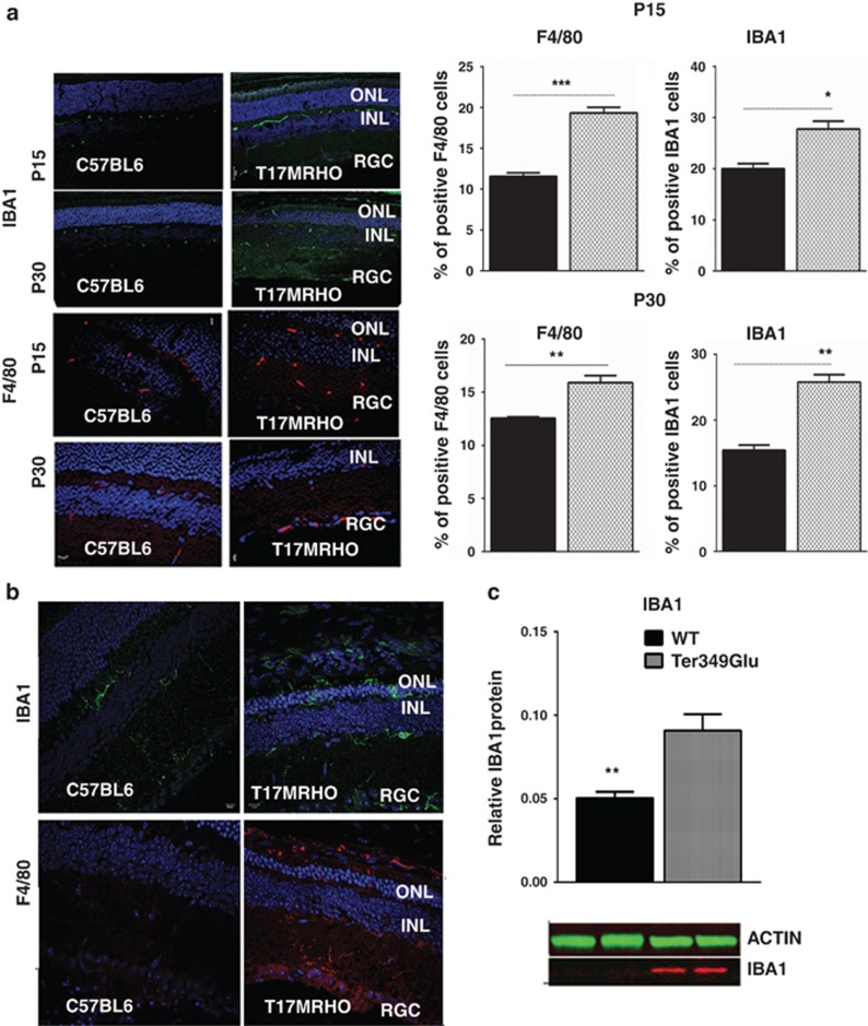
Recent studies on the endoplasmic reticulum stress have shown that the unfolded protein response (UPR) is involved in the pathogenesis of inherited retinal degeneration caused by mutant rhodopsin. However, the main question of whether UPR activation actually triggers retinal degeneration remains to be addressed. Thus, in this study, we created a mouse model for retinal degeneration caused by a persistently activated UPR to assess the physiological and morphological parameters associated with this disease state and to highlight a potential mechanism by which the UPR can promote retinal degeneration. We performed an intraocular injection in C57BL6 mice with a known unfolded protein response (UPR) inducer, tunicamycin (Tn) and examined animals by electroretinography (ERG), spectral domain optical coherence tomography (SD-OCT) and histological analyses. We detected a significant loss of photoreceptor function (over 60%) and retinal structure (35%) 30 days post treatment. Analysis of retinal protein extracts demonstrated a significant upregulation of inflammatory markers including interleukin-1β (IL-1β), IL-6, tumor necrosis factor-α (TNF-α), monocyte chemoattractant protein-1 (MCP-1) and IBA1. Similarly, we detected a strong inflammatory response in mice expressing either Ter349Glu or T17M rhodopsin (RHO). These mutant rhodopsin species induce severe retinal degeneration and T17M rhodopsin elicits UPR activation when expressed in mice. RNA and protein analysis revealed a significant upregulation of pro- and anti-inflammatory markers such as IL-1β, IL-6, p65 nuclear factor kappa B (NF-kB) and MCP-1, as well as activation of F4/80 and IBA1 microglial markers in both the retinas expressing mutant rhodopsins. We then assessed if the Tn-induced inflammatory marker IL-1β was capable of inducing retinal degeneration by injecting C57BL6 mice with a recombinant IL-1β. We observed ~19% reduction in ERG a-wave amplitudes and a 29% loss of photoreceptor cells compared with control retinas, suggesting a potential link between pro-inflammatory cytokines and retinal pathophysiological effects. Our work demonstrates that in the context of an established animal model for ocular disease, the persistent activation of the UPR could be responsible for promoting retinal degeneration via the UPR-induced pro-inflammatory cytokine IL-1β.


Figures

Figure 1
Figure 1
Persistently activated UPR in the wild-type retina induces retinal degeneration. The distribution of data values is shown in S.E.M. (a) Western blot analysis of Tn- or PBS-injected retinal protein extracts (N=4). Upper: a dose of 0.01 mg Tn activated the UPR in the retina 3 days post treatment. The UPR markers pEIF2a, CHOP and pATF6 were significantly increased compared with PBS-injected retinas (P=0.001, P=0.028 and P=0.020, respectively). Images of western blots are shown on the side. Bottom panel: activation of the UPR was observed concomitantly with the induction of inflammatory signaling in Tn-injected wild-type retinas. The inflammation markers IL-1β, IL-6, MCP-1 and TNF were upregulated 3 days post injection, suggesting that Tn injection induced not only UPR activation but also led to activation of an inflammatory response in the retina (P=0.001, P=0.029, P=0.021 and P=0.002). The calculation of the Tn-induced microglial response is shown in Supplementary Figure S1. (b) Scotopic ERG responses were significantly reduced in Tn-injected retinas at 10 and 30 days after treatment (N=6). Although no difference was observed between the PBS-injected and naive retinas, the a- and b-wave amplitudes were reduced by >60%, 30 days after Tn treatment (*P<0.05, **P<0.01, ***P<0.001 and ****P<0.0001). (c) A loss of retinal integrity in Tn-injected wild-type retinas was measured by SD-OCT (N=6). A >30% reduction in the ONL thickness was observed across all of the Tn-injected retina compared with PBS-injected mice, ****P<0.0001 at all measurement points. Bottom: SD-OCT images taken from PBS- and Tn-injected retinas. (d) Histological analysis following H&E staining of Tn-injected cryostat-sectioned retinas demonstrated a significant loss of photoreceptor cells. The number of rows of photoreceptor nuclei in the Tn-injected retinas was 36% lower compared with PBS-injected mice. Bottom: images of the H&E-stained retinas 30 days after treatment, (P=0.001). Scale bar indicates 20 μm
Figure 2
Figure 2
Injection with Tn leads to over production of cytokines in the retinal cells. (a) The cone-derived 661W cells treated with Tn (N=4) were harvested 1 and 8 h post injection to assess levels of Il-1β, Il-1 R and Il-6 by qRT-PCR. Results of the experiments demonstrated that the Tn treatment induces a 3.6-fold upregulation of Il-1β mRNA and 33% downregulation of Il-1R mRNA 1 h post treatment. (a) A 4-fold overexpression of Il-6 mRNA was observed 8 h post treatment with Tn. (b–d): Modulation of the UPR markers leads to altered cytokine's production. (b) Images of western blots obtained from the CHOP−/− and ATF4 overexpressing retinal extracts. (c) CHOP ablation in Tn-injected retinas (N=4) leads to a 46% and by 66% reduction in IL-6 and IL-1β, respectively, at 3 days post treatment, indicating that CHOP might regulate production of these cytokines. (d) The 2.6-fold increase in ATF4 triggers pro-inflammatory IL-1β over production. A 3-fold increase in IL-1β in AAV2/5 ATF4-injected retinas (N=4), suggested that the PERK UPR arm that leads to the ATF4 mRNA increase may be responsible for activation of the IL-1β mediated inflammatory signaling. The distribution of data values is shown in S.E.M., *P<0.05, **P<0.01, ***P<0.001 and ****P<0. 0001
Figure 3
Figure 3
ADRP progression in T17M RHO mice was accompanied by activation of the inflammatory response as measured by qRT-PCR (N=4). Pro- and anti-inflammatory markers were detected in P15, 30, 45 and 60 ADRP retinas. The distribution of data values is shown in S.E.M., *P<0.05, **P<0.01, ***P<0.001 and ****P<0. 0001
Figure 4
Figure 4
ADRP progression in T17M RHO mice is accompanied by activation of the inflammatory response as measured by western blot analysis (N=4). IL-1β, IL-6, p65 NF-Kβ, MCP-1 and TNF are significantly upregulated in the P15 ADRP retina (P=0.004, P=0.002, P=0.012, P=0.037 and P=0.002, respectively). Images of the western blots are shown on the side
Figure 5
Figure 5
An activated UPR was found in ADRP retinas expressing a class I Ter349Glu RHO mutant (N=4). The distribution of data values is shown in S.E.M. Upper panel: expression of UPR-associated genes was elevated in Ter349Glu RHO retinas during ADRP progression as measured by qRT-PCR. Mainly, overexpression of UPR-associated genes was detected at P30 (*P<0.05, **P<0.01, ***P<0.001 and ****P<0. 0001). Bottom panel: western blot analysis revealed activation of UPR markers CHOP and pATF6 (P=0.020 for both) (N=4). Images of the western blots are shown on the side. In addition, the inflammatory marker IL-1β was found to be increased by 1.9-fold in P30 Ter349Glu retinas (P=0.020)
Figure 6
Figure 6
Microglia are activated during ADRP progression. (a) The microglial markers F4/80 and IBA1 were used to perform immunohistochemical analysis in P15 and P30 cryostat-sectioned T17M RHO retinas (N=4). Five images of each individual T17M RHO and C57BL6 retina were taken to count positive cells. The number of positive cells was used to plot graphs for statistical analysis. The observed increase in F4/80-positive cells was 67% and 26%, respectively (P=0.01 for both time points). In addition, there was an increase in IBA-1 positive cells during ADRP progression from 40–70% compared with controls (P=0.013 and P=0.002, respectively). The distribution of data values are shown in S.E.M. Representative confocal images of P15 and P30 retinas are shown. (b) Activation of the microglial markers IBA1 and F4/80 were found in 10-week-old Ter349Glu retinas. Representative images are shown. (c) Almost 2-fold increase in IBA1 protein was found in Ter349Glu retinal protein extracts, P=0.014 (N=4). Bottom: images of the western blot treated with anti-Iba1 and anti-β-actin antibodies
Figure 7
Figure 7
Overexpression of the recombinant IL-1β cytokine can induce retinal degeneration in the wild-type retina. (a) Intraocular injection of 250 ng of recombinant IL-1β led to a statistically significant loss of scotopic ERG amplitudes 30 days after treatment (N=6). The a-wave and b-wave amplitudes were significantly reduced (P=0.035 and P=0.037, respectively). (b) SD-OCT analysis confirmed the ERG data indicating a >10% loss of the average ONL thickness in the superior and inferior regions of the IL-1β-injected retina (P=0.007 and P=0.0004, respectively) (N=4). Bottom: SD-OCT images of the PBS- and IL-1β-injected retinas. (c) Histological analysis following H&E staining of cryostat-sectioned IL-1β-injected retinas revealed a 29% loss of photoreceptor cells as compared with PBS-injected mice 30 days after treatment (P=0.001). Images of H&E stained PBS- and IL-1β-injected retinas are shown on the side. Scale bar indicates 20 μm

References

    1. 1Gorbatyuk M, Gorbatyuk O. Review: retinal degeneration: focus on the unfolded protein response. Mol Vis 2013; 19: 1995–1998. - PMC - PubMed
    1. 2Walter P, Ron D. The unfolded protein response: from stress pathway to homeostatic regulation. Science 2011; 334: 1081–1086. - PubMed
    1. 3Li GY, Fan B, Jiao YY. Rapamycin attenuates visible light-induced injury in retinal photoreceptor cells via inhibiting endoplasmic reticulum stress. Brain Res 2014; 1563: 1–12. - PubMed
    1. 4Wang T, Chen J. Induction of the unfolded-protein response by constitutive g-protein signaling in rod photoreceptor cells. J Biol Chem 2014; 289: 29310–29321. - PMC - PubMed
    1. 5Gorbatyuk MS, Gorbatyuk OS, LaVail MM, Lin JH, Hauswirth WW, Lewin AS. Functional rescue of P23H rhodopsin photoreceptors by gene delivery. Adv Exp Med Biol 2012; 723: 191–197. - PMC - PubMed

Publication types

MeSH terms