. 2014 Oct 31;18(3):pyu074.
doi: 10.1093/ijnp/pyu074.

Strain-dependent variations in stress coping behavior are mediated by a 5-HT/GABA interaction within the prefrontal corticolimbic system

Affiliations

Strain-dependent variations in stress coping behavior are mediated by a 5-HT/GABA interaction within the prefrontal corticolimbic system

Diego Andolina et al. Int J Neuropsychopharmacol. .

Abstract

Background: Serotonin and γ-aminobutyric acid (GABA) transmission is crucial in coping strategies.

Methods: Here, using mice from 2 inbred strains widely exploited in behavioral neurochemistry, we investigated whether serotonin transmission in medial prefrontal cortex and GABA in basolateral amygdala determine strain-dependent liability to stress response and differences in coping.

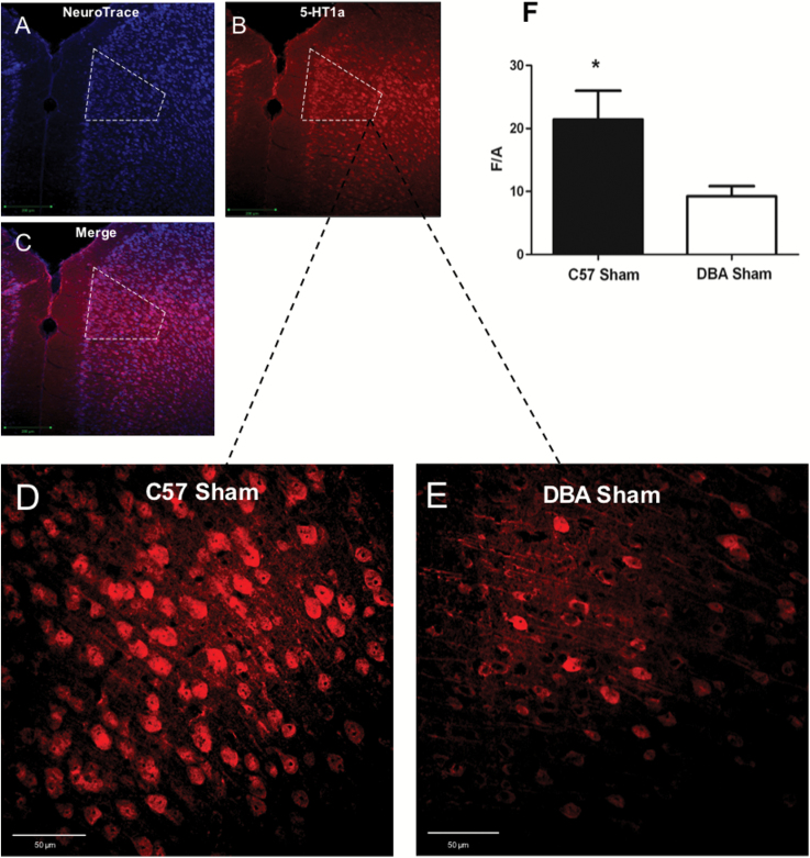
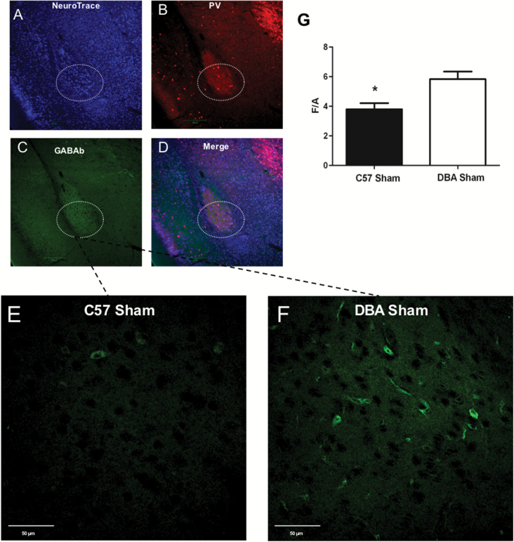
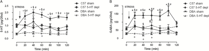
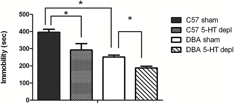
Results: C57BL/6J mice displayed greater immobility in the forced swimming test, higher serotonin outflow in medial prefrontal cortex, higher GABA outflow in basolateral amygdala induced by stress, and higher serotonin 1A receptor levels in medial prefrontal cortex accompanied by lower GABAb receptor levels in basolateral amygdala than DBA/2J mice. In assessing whether serotonin in medial prefrontal cortex determines GABA functioning in response to stress and passive coping behavior in C57BL/6J and DBA/2J mice, we observed that selective prefrontal serotonin depletion in C57BL/6J and DBA/2J reduced stress-induced GABA outflow in basolateral amygdala and immobility in the forced swimming test.

Conclusions: These results show that strain-dependent prefrontal corticolimbic serotonin/GABA regulation determines the strain differences in stress-coping behavior in the forced swimming test and point to a role of a specific neuronal system in genetic susceptibility to stress that opens up new prospects for innovative therapies for stress disorders.

Keywords: GABA; basolateral amygdala; medial prefrontal cortex; serotonin; strain.


Figures

Figure 1.
Figure 1.
Representative positions of approximate location of 5,7-dihydroxytryptamine (5,7-DHT) point microinjection (white dot) in medial prefrontal cortex (mpFC) in C57BL/6J (C57) and DBA/2J (DBA) (A). The arrow indicates the point of microinjection. Representative positions of the probe in the mpFC (B) and basolateral amygdala (BLA) (C) in DBA mice and C57 mice. The segment of the membrane probe is also shown.
Figure 2.
Figure 2.
Serotonin (5-HT1A) immunoreactivity in the medial prefrontal cortex (mpFC) prelimbic area (PL) of C57BL/6J (C57) sham and DBA/2J (DBA) sham double-labeled confocal images of NeuroTrace counterstaining (A: blue) and 5-HT1A labeling (B: red) plus merged (C). Panels D and E are higher magnification pictures from PL of C57 sham (D) and DBA sham (E) reacted with 5-HT1A antibody. Histogram of densitometric values (F) of 5-HT1A immunofluorescence expressed as mean fluorescence of individual cells normalized to total cellular surface (F/A). Data are reported as means ± SD (n=6 animal/group). *P<.05; scale bars: A-C=200 μm; D-E=50 μm.
Figure 3.
Figure 3.
GABAb immunoreactivity in the basolateral amygdala (BLA) of C57BL/6J (C57) sham and DBA/2J (DBA) sham. Triple-labeled confocal images of NeuroTrace counterstaining (A: blue), and parvalbumin labeling (B: red), GABAb (C: green) plus merged (D) of BLA. Panels E and F are higher magnification pictures from BLA of C57 (E) and DBA (F) reacted with GABAb antibody. Histogram of densitometric values (G) of GABAb immunofluorescence expressed as mean fluorescence of individual cells normalized to total cellular surface (F/A). Data are reported as means ± SD (n=6 animals/group). *P<.05. Scale bars: A-D=200 μm; E-F=50 μm.
Figure 4.
Figure 4.
Effects of 120-minute restraint stress on serotonin (5-HT) outflow in the medial prefrontal cortex (mpFC) (A) and GABA outflow in the basolateral amygdala (BLA) (B) of sham C57 and DBA mice and of C57 and DBA mice bearing a selective prefrontal cortical 5-HT depletion in the mpFC (C57 5-HT Depl, DBA 5-HT Depl). Arrows indicate the beginning of the restraint. §P<.05 from the basal values. *P<.05, C57 sham in comparison with the corresponding time point of DBA sham group. #P<.05 C57 sham in comparison with the corresponding time point of C57 5-HT Depl group. +P<.05 DBA sham in comparison with the corresponding time point of DBA 5-HT Depl group.
Figure 5.
Figure 5.
Effects of strain (C57 sham, DBA sham) and of selective prefrontal cortical serotonin (5-HT) depletion in the medial prefrontal cortex (mpFC) of C57 and DBA mice (C57 5-HT Depl; DBA 5-HT Depl) on immobility in the forced swimming test (FST). Results are expressed as mean ± SE duration (second) of immobility. *P<.05. C57 sham in comparison with DBA sham group, C57 5-HT-Depl group and DBA sham group in comparison with DBA 5-HT-Depl group.
Figure 6.
Figure 6.
Effects of strain (C57 sham, DBA sham) and of selective prefrontal cortical serotonin (5-HT) depletion in the medial prefrontal cortex (mpFC) of C57 and DBA mice (C57 5-HT Depl; DBA 5-HT Depl) on anxiety behavior in the elevated plus maze. Data are expressed as mean percent time spent or mean percent entries made in the open arm and as mean percent time spent or mean percent entries made in closed arms ± SE. *P<.05.
Figure 7.
Figure 7.
Effects of strain (C57 sham, DBA sham) and of selective prefrontal cortical serotonin (5-HT) depletion in the medial prefrontal cortex (mpFC) of C57 and DBA mice (C57 5-HT Depl; DBA 5-HT Depl) on locomotor activity in the open field. Results are expressed as mean ± SE distance moved (cm).

References

    1. Ago Y, Koyama Y, Baba A, Matsuda T. (2003). Regulation by 5-HT1A receptors of the in vivo release of 5-HT and DA in mouse frontal cortex. Neuropharmacology 45:1050–1056. - PubMed
    1. Akirav I, Maroun M. (2007). The role of the medial prefrontal cortex-amygdala circuit in stress effects on the extinction of fear. Neural Plast 2007:30873. - PMC - PubMed
    1. Albert PR, Zhou QY, Van Tol HH, Bunzow JR, Civelli O. (1990). Cloning, functional expression, and mRNA tissue distribution of the rat 5-hydroxytryptamine1A receptor gene. J Biol Chem 265:5825–5832. - PubMed
    1. Alcaro A, Cabib S, Ventura R, Puglisi-Allegra S. (2002). Genotype and experience dependent susceptibility to depressive-like responses in the forced-swimming test. Psychopharmacology (Berl) 164:138–143. - PubMed
    1. Amat J, Matus-Amat P, Watkins LR, Maier SF. (1998). Escapable and inescapable stress differentially alter extracellular levels of 5-HT in the basolateral amygdala of the rat. Brain Res 812:113–120. - PubMed

Publication types

MeSH terms