Review
. 2014 Dec 19:9:32.
doi: 10.1186/s13062-014-0032-7.

Once upon a time the cell membranes: 175 years of cell boundary research

Affiliations
Review

Once upon a time the cell membranes: 175 years of cell boundary research

Jonathan Lombard. Biol Direct. .

Abstract

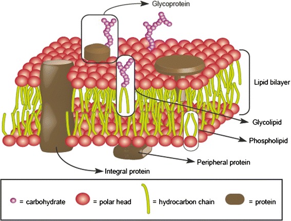
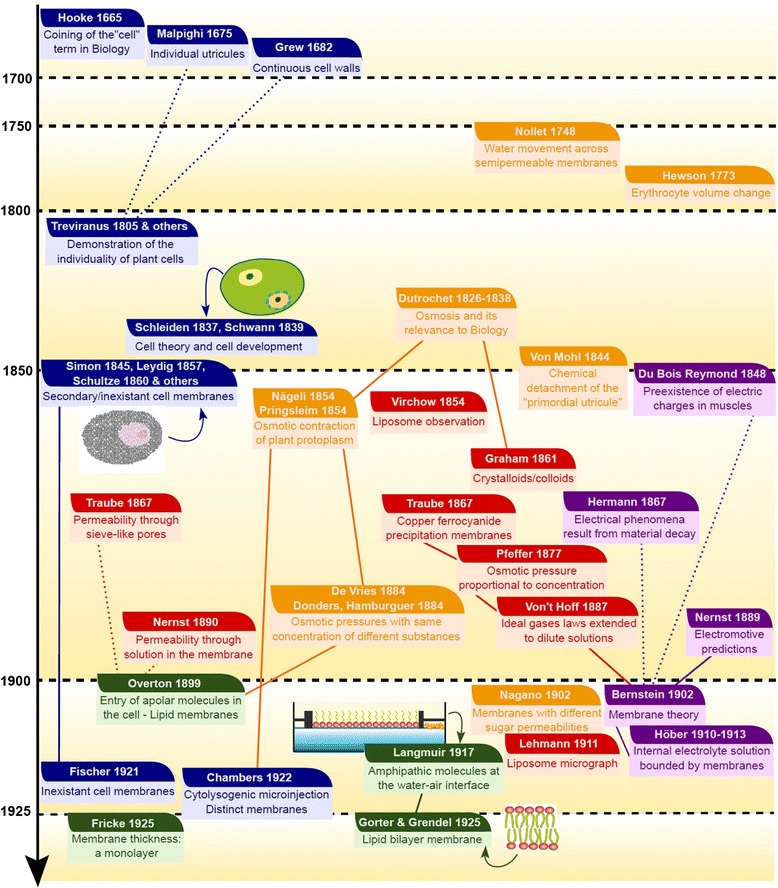
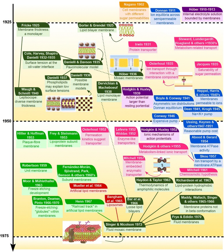
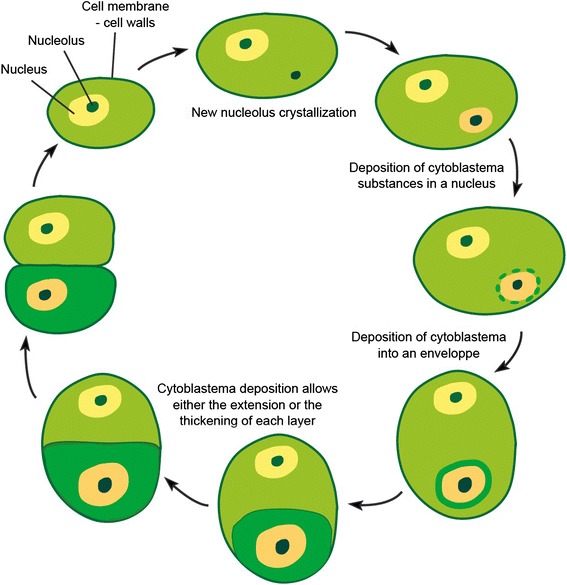
All modern cells are bounded by cell membranes best described by the fluid mosaic model. This statement is so widely accepted by biologists that little attention is generally given to the theoretical importance of cell membranes in describing the cell. This has not always been the case. When the Cell Theory was first formulated in the XIX(th) century, almost nothing was known about the cell membranes. It was not until well into the XX(th) century that the existence of the plasma membrane was broadly accepted and, even then, the fluid mosaic model did not prevail until the 1970s. How were the cell boundaries considered between the articulation of the Cell Theory around 1839 and the formulation of the fluid mosaic model that has described the cell membranes since 1972? In this review I will summarize the major historical discoveries and theories that tackled the existence and structure of membranes and I will analyze how these theories impacted the understanding of the cell. Apart from its purely historical relevance, this account can provide a starting point for considering the theoretical significance of membranes to the definition of the cell and could have implications for research on early life.


Figures

Figure 1
Figure 1
Fluid mosaic model. Schematic view of biological membrane structure as currently depicted.
Figure 2
Figure 2
Timeline 1665–1925. Summary of the main contributions related to cell membrane discovery between the coining of the term “cell” in biology and the first studies on cell membrane structures. The events are approximatively ordered from top to bottom from the earlier events to the most recent. Although the studies are sometimes difficult to classify, the colors of the boxes reflect some major research axes influent to this history: dark blue, doubts about the existence of cell membranes; orange, osmotic studies; red, studies with artificial membranes; purple, electrophysiology works; dark green, direct description of membranes. Although most of these contributions were highly interconnected, full lines between boxes highlight particularly important relationships and dashed lines point out to contradictory views in major controversies.
Figure 3
Figure 3
Timeline 1925–1972. Summary of the main contributions related to cell membrane discovery between the first studies on cell membrane structures and the formulation of the fluid mosaic model. The events are approximatively ordered from top to bottom from the earlier events to the most recent. Although the studies are sometimes difficult to classify, the colors of the boxes reflect some major research axes influent to this history: orange, osmotic studies; red, studies with artificial membranes; purple, electrophysiology works; dark green, direct description of membranes; pink, some transporter theories; light blue, asymmetric ion distribution debate; light green, electron microscopy studies. Although most of these contributions were highly interconnected, full lines between boxes highlight particularly important relationships and dashed lines point out to contradictory views in major controversies.
Figure 4
Figure 4
The development of cells according to Schleiden. This figure has been drawn for clarity from descriptions by Schleiden and Schwann, but these authors never tried to provide such a synthetic depiction in their work. Schwann’s model was very similar, except for his opinion that new cells could also crystallize from cytoblastema outside previous cells.
Figure 5
Figure 5
XIX th century doubts about the existence of membranes. A. In this vision, the cell is devoid of any membrane and all the properties of the cell are defined by the activity of the protoplasmic colloid. B. The cell is surrounded by an external layer (membrane) of which the nature is distinct to the rest of the protoplasm. Yet, in this view, the inside of the cell remains a colloid.
Figure 6
Figure 6
Oil at air/water interfaces. A. Oil molecules spontaneously spread on the air/water interface until they form a layer one molecule thick. B. The Langmuir trough allows to precisely measure the surface that these monolayers can spread depending on the applied pressure.
Figure 7
Figure 7
Surface measurement of membrane lipid monolayers as a way to determine membrane structure. A. Summary of the method, consisting in the comparison between the surface occupied by lipids extracted from membranes and the estimated surface of cells B. Different results and interpretations.
Figure 8
Figure 8
Membrane structure hypotheses in the 1930’s. A. Paucimolecular model, with a lipid bilayer coated with proteins in both sides. B. Höber’s mosaic model in which membranes behaved both as solvents and sieves.
Figure 9
Figure 9
Possible molecular arrangements of biological membranes redrawn from Danielli in 1936 [ 101 ] . A-E. Cross-section of hypothetical membranes with internal lipids and coating proteins. F. Cross-section of an hypothetical membrane made up of lipoprotein subunits. G-I. Surface of mosaic membranes.
Figure 10
Figure 10
Donnan’s equilibrium. Two solutions containing two different initial concentrations of different salts are separated by a membrane. In this case, the membrane is impermeable to anions but permeable to cations. Donnan thermodynamic calculations and experiments showed that, contrary to what could be initially thought, the two cations do not just interchange with each other until they are equally distributed in the two compartments. Instead, equivalent quantities of both cations cross the membrane; as their initial concentrations are different, the cation which was initially less concentrated proportionally crosses the membrane more than the initially highly concentrated cation.
Figure 11
Figure 11
Redrawings of some examples of transport across membranes in the 1950’s and 1960’s. A. Eccles depicts in 1963 the coupling between a metabolic-driven ion pump and several different channels [165]. B. Burgen suggests in 1957 that molecules cross pores thanks to specific and dynamic interactions with them [166]. C. Mitchell describes in 1957 an enzymatic-like protein transporter embedded in the membrane [167]. D. Mitchell’s chemiosmotic hypothesis in 1961 is based in the existence of structures embedded in the biological membranes [168]. E. From an early date, Danielli and collaborators considered the possibility that channels may have existed within their paucimolecular hypothesis (redrawn from [169]). F. Danielli’s summary of different transporter models in 1954 (redrawn from [134]).

References

    1. Singer SJ, Nicolson GL. The fluid mosaic model of the structure of cell membranes. Science. 1972;175:720–731. doi: 10.1126/science.175.4023.720. - DOI - PubMed
    1. Tanford C. The Hydrophobic Effect. Formation of Micelles and Biological Membranes. New York: Wiley; 1973.
    1. Daleke DL, Lyles JV. Identification and purification of aminophopholipid flippases. Biochem Biophys Acta. 2000;1486:108–127. - PubMed
    1. Ling GN. Debunking the alleged resurrection of the sodium pump hypothesis. Physiol Chem Phys Med NMR. 1997;29:123–198. - PubMed
    1. Edidin M. Lipids on the frontier: a century of cell-membrane bilayers. Nat Rev Mol Cell Biol. 2003;4:414–418. doi: 10.1038/nrm1102. - DOI - PubMed

Publication types

LinkOut - more resources