Review
. 2014 Dec 19:9:144.
doi: 10.1186/s13023-014-0144-2.

Ventricular septal defect

Affiliations
Review

Ventricular septal defect

Diane E Spicer et al. Orphanet J Rare Dis. .

Abstract

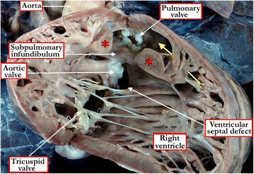
Background: Ventricular septal defects are the commonest congenital cardiac malformations. They can exist in isolation, but are also found as integral components of other cardiac anomalies, such as tetralogy of Fallot, double outlet right ventricle, or common arterial trunk. As yet, there is no agreement on how best to classify such defects, nor even on the curved surface that is taken to represent the defect.

Methods: Based on our previous pathological and clinical experiences, we have reviewed the history of classification of holes between the ventricles. We proposed that the defects are best defined as representing the area of deficient ventricular septation. This then permits the recognition of clinically significant variants according to the anatomic borders, and the way the curved surface representing the area of deficient septation opens into the morphologically right ventricle.

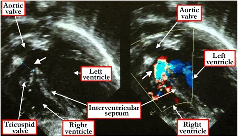
Results: Clinical manifestation depends on the size of the defect, and on the relationship between systemic and pulmonary vascular resistances. Symptoms include failure to thrive, along with the manifestations of the increase in flow of blood to the lungs. Diagnosis can be made by physical examination, but is confirmed by echocardiographic interrogation, which delineates the precise anatomy, and also provides the physiologic information required for optimal clinical decision-making. Cardiac catheterization offers additional information regarding hemodynamics, particularly if there is a concern regarding an increase in pulmonary vascular resistance. Hemodynamic assessment is rarely necessary to make decisions regarding management, although it can be helpful if assessing symptomatic adults with hemodynamically restrictive defects. In infants with defects producing large shunts, surgical closure is now recommended in most instances as soon as symptoms manifest. Only in rare cases is palliative banding of the pulmonary trunk now recommended. Closure with devices inserted on catheters is now the preferred approach for many patients with muscular defects, often using a hybrid procedure. Therapeutic closure should now be anticipated with virtually zero mortality, and with excellent anticipated long-term survival.

Conclusion: Ventricular septal defects are best defined as representing the borders of the area of deficient ventricular septation. An approach on this basis permits recognition of the clinically significant phenotypic variants.


Figures

Figure 1
Figure 1
The images show the problems existing in defining the boundaries of the area of space that represents the “ventricular septal defect”. Panel A shows a simulated five chamber echocardiographic cut in a specimen with overriding of the aortic root relative to the apical muscular septum. The yellow arrow shows the continuation of the long axis of the muscular ventricular septum. This area marks the true geometric interventricular communication, which is almost planar. This virtual plane, however, can never be closed, since its cranial margin is formed by the leaflets of overriding aortic root. The red double-headed arrow shows the margins of the curved surface that would be closed so as to restore septal integrity. Although shown as a planar entity, in reality the surface is markedly curved due to the non-planar configurations of its boundaries. It is shown in planar format for the sake of simplicity. As shown in Panel B, it is the margins of this curved surface, outlined by the red dots, that are taken as representing the ventricular septal defect when viewed from the right ventricle. Note that its cranial border is formed by the muscular outlet septum, which is malaligned relative to the apical muscular septum, accentuating the non-planar configuration of its surface.
Figure 2
Figure 2
These images show that the interventricular communication is not necessarily the same thing as the ventricular septal defect. In Figure 2 A, we show a heart with double outlet right ventricle sectioned in four-chamber fashion, showing the aorta arising exclusively from the right ventricle, but with its cranial margin formed by fibrous continuity between the leaflets of the aortic and mitral valves. It is the space between this margin and the crest of the apical muscular septum that is the true interventricular communication. This space (double headed red arrow), however, can never be closed, since such closure would wall off the aorta from the left ventricle. As shown in Panel B, in which the free wall of the right ventricle has been lifted away to reveal a defect in a heart with the larger part of the aortic root supported within the right ventricle, in other words effectively a double outlet ventriculo-arterial connection, the outlet septum is exclusively a right ventricular structure, and is fibrous rather than muscular. The yellow dots show the margins of the defect that would be closed so as to place the aortic root in continuity with the cavity of the left ventricle. It is this curved surface that represents the ventricular septal defect, albeit that it is not the geometric interventricular communication.
Figure 3
Figure 3
In this heart, the right ventricle has been windowed, and the structures are viewed from the ventricular apex. As also seen in Figure 2B, the aortic root overrides the crest of the apical muscular septum, and the greater part of the aortic root is supported within the right ventricle, with the antero-cephalad deviation of the muscular outlet septum producing subpulmonary infundibular stenosis. The specimen shows the tetralogy variant of hearts with double outlet from the right ventricle, but the red dots show the boundaries of the curved surface that is taken to represent the ventricular septal defect.
Figure 4
Figure 4
The view of the opened right atrioventricular junction (A) shows a muscular ventricular septal defect opening to the inlet of the right ventricle beneath the septal leaflet of the tricuspid valve. In the specimen shown in Figure 4 B, the free wall of the right ventricle has been lifted away to show a muscular ventricular septal defect opening into the outlet portion of the right ventricle. The ventricular septal defect lies within the arms of the septal band (red Y), with the caudal arm fusing with the inner heart curvature to produce a muscular bar (yellow dots) that interposes between the leaflets of the atrioventricular and arterial valves.
Figure 5
Figure 5
The right ventricle has been windowed in this heart to show a perimembranous ventricular septal defect opening to the outlet of the right ventricle, with malalignment of the muscular outlet septum so that a small part of the aortic root overrides the crest of the muscular ventricular septum. The yellow dots mark the fibrous continuity between the leaflets of the tricuspid and the aortic valves, the feature which marks the defect as being perimembranous. Note that, despite the anterior deviation of the muscular outlet septum, which forms the cranial margin of the defect, there is no subpulmonary stenosis. This is an example of the so-called Eisenmenger defect.
Figure 6
Figure 6
In this heart (A) with a doubly committed and juxta-arterial ventricular septal defect there is fibrous continuity between the leaflets of the arterial valves (black dots) at the roof of the defect, with a muscular bar (yellow dots) separating the leaflets of the tricuspid and aortic valves. The muscular bar is formed by fusion of the caudal limb of the septal band (red Y) with the inner heart curvature, the latter also known as the ventriculo-infundibular fold. As shown in Figure 6 B, defects with fibrous continuity between the leaflets of the arterial valves (black dots), due to failure of muscularisation of the subpulmonary infundibulum, can also extend to become perimembranous. The leaflets of the tricuspid and aortic valves in this specimen are in fibrous continuity (yellow dots), with the defect again positioned within the Y of the septal band.
Figure 7
Figure 7
The view from the left ventricle in this specimen shows a ventricular septal defect with exclusively muscular borders opening towards the outlet of the right ventricle, but with postero-caudal deviation of the muscular outlet septum (yellow dots), causing subaortic stenosis.
Figure 8
Figure 8
As shown in these specimens, it is crucial to identify the borders of ventricular septal defects, since this provides information regarding the location of the atrioventricular conduction axis. As shown in Figure 8 A, the axis (red dots) will run along the postero-inferior aspect of perimembranous ventricular septal defects, in other words to the right hand of the surgeon approaching through the tricuspid valve. In contrast, when a muscular defect opens to the inlet of the right ventricle, as shown in Figure 8 B, the conduction axis is located to the left hand of the surgeon approaching through the tricuspid valve. The red star shows the location of the atrioventricular node at the apex of the triangle of Koch (yellow lines).
Figure 9
Figure 9
The rule regarding the location of the conduction axis (red dots) in hearts with perimembranous ventricular septal defects holds good for all instances except when there is malalignment between the atrial septum and the apical muscular septum. This is associated with straddling and overriding of the tricuspid valve. As shown in the specimen, the conduction axis, carried on the crest of the muscular septum, can no longer take origin from the regular atrioventricular node, which remains at the apex of the triangle of Koch (yellow lines). Instead, it originates from an anomalous node (red star), which is formed at the site of union between the malaligned muscular septum and the inferior atrioventricular junction.
Figure 10
Figure 10
Defects can also open to the inlet of the right ventricle when they are part of an atrioventricular septal defect with a common atrioventricular valve. As shown in Figure 10 A, when the superior and inferior bridging leaflets are attached to the leading edge of the atrial septum (yellow dots) in this setting, then shunting is possible only at the ventricular level. The ventricular component of the defect (red dots) is then the true ventricular septal defect of atrioventricular canal type. Figure 10 B shows the left ventricular view of the same heart, with both bridging leaflets attached to the leading edge of the atrial septum (yellow dots). Note the atrial septal defect within the oval fossa, and the coronary sinus in the left atrioventricular groove.
Figure 11
Figure 11
The chest x-ray, viewed in the antero-posterior projection, from a typical patient with ventricular septal defect shows cardiomegaly, increased pulmonary vascular markings, and atelectasis of the left lower lobe.
Figure 12
Figure 12
The electrocardiogram, again from a typical patient with a ventricular septal defect, shows features of biventricular hypertrophy.
Figure 13
Figure 13
The cross-sectional echocardiogram, taken in slightly off axis four chamber projections, shows an apical muscular ventricular septal defect (white arrow) in the left hand panel of Figure 13 a. The right hand panel shows the Doppler color flow map of a hemodynamically restrictive muscular ventricular septal defect. The apical five chamber view, shown in Figure 13 b, shows the Doppler color flow map of an apical muscular ventricular septal defect (white arrow).
Figure 14
Figure 14
The subxyphoid four chamber cross-sectional echocardiogram shows the fibrous continuity between the leaflets of the aortic and tricuspid valves that reveals the defect to be perimembranous (white arrow) and opening to the inlet of the right ventricle (left hand panel). The right hand panel shows the color Doppler flow demonstrating the right-to-left shunt.
Figure 15
Figure 15
The short axis echocardiographic cut (left panel) demonstrates the fibrous continuity between the leaflets of the aortic and pulmonary valves that identifies the defect as being doubly committed and juxta-arterial. The right panel shows the Doppler color flow map of the same defect.
Figure 16
Figure 16
The panels show subcostal four chamber echocardiographic cuts revealing malalignment between the atrial and muscular ventricular septal structures in a heart with straddling and overriding of the tricuspid valve. Note the tendinous cords attached to a papillary muscle (yellow asterisk) within the left ventricle. The left hand panel shows the diastolic, and the right panel the systolic location of the overriding leaflets.
Figure 17
Figure 17
The three dimensional echocardiographic dataset, shown in four chamber fashion (A), reveals abnormal muscle bundles in the right ventricle (white arrows), producing so-called double chambered right ventricle. The Doppler color flow map in the same heart (B) confirms the presence of the double chambered right ventricle.
Figure 18
Figure 18
The free wall of the right ventricle has been lifted from the septal surface to show the features of tetralogy of Fallot with double chambered right ventricle due to anomalous apical septoparietal trabeculations (red asterisks). The ventricular septal defect is perimembranous. The yellow arrow marks the exit from the apical portion of the right ventricle to the subpulmonary outflow tract. Note the dysplastic and rudimentary leaflets of the pulmonary valve.

References

    1. Roberts WC. The 2 most common congenital heart diseases [Editorial] Am J Cardiol. 1984;53:1198. doi: 10.1016/0002-9149(84)90662-3. - DOI - PubMed
    1. Mitchell SC, Korones SB, Berendes HW. Congenital heart disease in 56,109 births. Incidence and natural history. Circulation. 1971;43:323–332. doi: 10.1161/01.CIR.43.3.323. - DOI - PubMed
    1. Soto B, Becker AE, Moulaert AJ, Lie JT, Anderson RH. Classification of ventricular septal defects. Br Heart J. 1980;43:332–343. doi: 10.1136/hrt.43.3.332. - DOI - PMC - PubMed
    1. Capelli H, Andrade JL, Somerville J. Classification of the site of ventricular septal defect by 2-dimensional echocardiography. Am J Cardiol. 1983;51:1474–1480. doi: 10.1016/0002-9149(83)90660-4. - DOI - PubMed
    1. Jacobs JP, Burke RP, Quintessenza JA, Mavroudis C. Congenital Heart Surgery Nomenclature and Database Project: ventricular septal defect. Ann Thor Surg. 2000;69(Suppl 4):S25–S35. doi: 10.1016/S0003-4975(99)01270-9. - DOI - PubMed

MeSH terms

LinkOut - more resources