

Quantification of global cerebral atrophy in multiple sclerosis from 3T MRI using SPM: the role of misclassification errors
- PMID: 25523616
- PMCID: PMC4409073
- DOI: 10.1111/jon.12194
Quantification of global cerebral atrophy in multiple sclerosis from 3T MRI using SPM: the role of misclassification errors
Abstract
Purpose: We tested the validity of a freely available segmentation pipeline to measure compartmental brain volumes from 3T MRI in patients with multiple sclerosis (MS). Our primary focus was methodological to explore the effect of segmentation corrections on the clinical relevance of the output metrics.
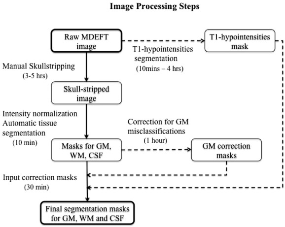
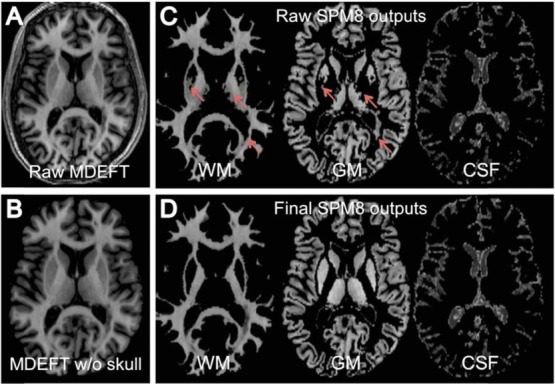
Methods: Three-dimensional T1-weighted images were acquired to compare 61 MS patients to 30 age- and gender-matched normal controls (NC). We also tested the within patient MRI relationship to disability (eg, expanded disability status scale [EDSS] score) and cognition. Statistical parametric mapping v. 8 (SPM8)-derived gray matter (GMF), white matter (WMF), and total brain parenchyma fractions (BPF) were derived before and after correcting errors from T1 hypointense MS lesions and/or ineffective deep GM contouring.
Results: MS patients had lower GMF and BPF as compared to NC (P<.05). Cognitively impaired patients had lower BPF than cognitively preserved patients (P<.05). BPF was related to EDSS; BPF and GMF were related to disease duration (all P<.05). Errors caused bias in GMFs and WMFs but had no discernable influence on BPFs or any MRI-clinical associations.
Conclusions: We report the validity of a segmentation pipeline for the detection of MS-related brain atrophy with 3T MRI. Longitudinal studies are warranted to extend these results.
Keywords: Brain atrophy; MRI; gray matter; lesions; multiple sclerosis; segmentation.
© 2014 The Authors. Journal of Neuroimaging published by Wiley Periodicals, Inc. on behalf of the American Society of Neuroimaging.
Figures
References
-
- Bermel RA, Bakshi R. The measurement and clinical relevance of brain atrophy in multiple sclerosis. Lancet Neurol. 2006;5:158–170. - PubMed
-
- Sanfilipo MP, Benedict RH, Sharma J. The relationship between whole brain volume and disability in multiple sclerosis: a comparison of normalized gray vs. white matter with misclassification correction. NeuroImage. 2005;26:1068–1077. - PubMed
-
- Dalton CM, Chard DT, Davies GR. Early development of multiple sclerosis is associated with progressive grey matter atrophy in patients presenting with clinically isolated syndromes. Brain. 2004;127:1101–1107. - PubMed
-
- Pirko I, Lucchinetti CF, Sriram S. Gray matter involvement in multiple sclerosis. Neurology. 2007;68:634–642. - PubMed
MeSH terms
LinkOut - more resources
Full Text Sources
Other Literature Sources
Medical
