. 2015;7(1):15-25.
doi: 10.4161/19420862.2015.989032.

Structural insights and biomedical potential of IgNAR scaffolds from sharks

Affiliations

Structural insights and biomedical potential of IgNAR scaffolds from sharks

Stefan Zielonka et al. MAbs. 2015.

Abstract

In addition to antibodies with the classical composition of heavy and light chains, the adaptive immune repertoire of sharks also includes a heavy-chain only isotype, where antigen binding is mediated exclusively by a small and highly stable domain, referred to as vNAR. In recent years, due to their high affinity and specificity combined with their small size, high physicochemical stability and low-cost of production, vNAR fragments have evolved as promising target-binding scaffolds that can be tailor-made for applications in medicine and biotechnology. This review highlights the structural features of vNAR molecules, addresses aspects of their generation using immunization or in vitro high throughput screening methods and provides examples of therapeutic, diagnostic and other biotechnological applications.

Keywords: CDR, complementarity-determining region; HV, hypervariable region; IgNAR; IgNAR V domain, variable domain of IgNAR; IgNAR, immunoglobulin new antigen receptor; VH, variable domain of the heavy chain; VHH, variable domain of camelid heavy chain antibodies; VL, variable domain of the light chain; antibody technology; biologic therapeutic; heavy chain antibody; mAbs, monoclonal antibodies; scFv, single chain variable fragment; shark; single chain binding domain; vNAR, variable domain of IgNAR.


Figures

Figure 1.
Figure 1.
Structural features of intact IgG (A), camelid hclgG (B), and shark IgNAR (C) antibody formats shown as surface representation (top) as well as ribbon and schematic representations (bottom). Individual domains are colored as indicated in the schematic representation; hinge regions are colored yellow; glycans not shown. (A) IgG model is based on pdb entry 1IGT. (B) Model of hclgG was generated by molecular replacement based on pdb entries 1IGT (for Fc region) and 1IEH (for VHH domain). 1IEH was aligned to the CH1 domain of 1IGT with YASARA structure. After deletion of absent domains (VL, CL, VH, and CH1), the VHH section was connected to the Fc region via a short camelid hinge sequence (see Hamers-Casterman et al.). Then, a 2-step energy minimization using YASARA2 force field was conducted to yield the depicted structure. (C) Coordinates of intact IgNAR including the hypothetical structure of IgNAR C5 domain were generously provided by Prof. Dr. Michael Sattler and Dr. Janosch Hennig (see Feige et al.). Picture rendered with POV-Ray (www.povray.org/).
Figure 2.
Figure 2.
Comparison of VH (left; from pdb entry 1IGT) and vNAR (right, from pdb entry 2COQ) binding domains depicted as ribbon representation as well as an overlay of both structures (middle). CDR1 and CDR3 are shown in gray. Two β strands and CDR2 of the VH domain are highlighted in orange. These structural elements are absent in the vNAR domain which possesses HV2 and HV4 (both highlighted in blue), instead. Disulfide bonds are shown as yellow sticks. Picture rendered with POV-Ray (www.povray.org/).
Figure 3.
Figure 3.
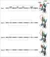
Different types of IgNAR V domains. Variable domains are categorized based on the presence or the absence of non-canonical cysteine residues (black dots). Canonical cysteine residues (white dots) and disulfide bonds (connecting lines), conserved tryptophan (W) as well as complementarity determining regions (CDR) and hypervariable loops (HV) are shown in their relative positions. Ribbon presentations of vNAR domains are depictions of pdb entries 1SQ22 (type I), 2COQ (type II), and 4HGK (type IV) as well as a modeled type III structure based on 2COQ. The latter was generated via homology modeling using YASARA structure. First, vNAR residues of 2COQ were changed to match a reported type III sequence (AAM76948 from Streltsov et al.). Then, side chain geometries were optimized followed by a 2-step energy minimization using the YASARA2 force field.
Figure 4.
Figure 4.
Translocon arrangements of immunoglobulin genes in higher vertebrates and cluster configuration of IgNAR genes of cartilaginous fish. In the translocon organization there are many variable (V) segments upstream of many diversity (D, only for heavy chains) and joining (J) segments that recombine randomly to encode the variable domain of the heavy chain or the light chain. IgNAR genes (like all Ig genes of the cartilaginous fish) are organized in the cluster configuration. Each cluster contains for the variable domain one V segment, 3 D segments and one J segment. Recombination occurs exclusively within one cluster. H, heavy chain loci; L, light chain loci, C, constant region.
Figure 5.
Figure 5.
Examples of CDR3 variability in vNAR domains depicted in ribbon representation. (A) Short loop (type IV, pdb entry 4HGK). (B) Large loop with one disulfide constraint (type II, pdb entry 2COQ). (C) Highly constrained loop tethered by 2 cystine motifs (type I, pdb entry 1SQ2). (D) Extended CDR3 forming an α- helical motif (type II, pdb entry 2I25). (E) Extended CDR3 forming a 2-stranded β-sheet (type IV, pdb entry 2Z8V). (F) Extended CDR3 incorporating an amyloid-β p3 fragment (type IV, pdb entry 3MOQ). (G) Overlay of structures A–F. Disulfide bonds are shown in yellow. Picture rendered with POV-Ray (www.povray.org/).

References

    1. Aggarwal RS. What's fueling the biotech engine-2012 to 2013. Nat Biotechnol 2014; 32:32-9; PMID:24406926; http://dx.doi.org/10.1038/nbt.2794 - DOI - PubMed
    1. Aggarwal SR. What's fueling the biotech engine-2011 to 2012. Nat Biotechnol 2012; 30:1191-7; PMID:23222785; http://dx.doi.org/10.1038/nbt.2437 - DOI - PubMed
    1. Buss NA, Henderson SJ, McFarlane M, Shenton JM, de Haan L. Monoclonal antibody therapeutics: history and future. Curr Opin Pharmacol 2012; 12:615-22; PMID:22920732; http://dx.doi.org/10.1016/j.coph.2012.08.001 - DOI - PubMed
    1. Reichert JM. Marketed therapeutic antibodies compendium. MAbs 2012; 4:413-5; PMID:22531442; http://dx.doi.org/10.4161/mabs.19931 - DOI - PMC - PubMed
    1. Reichert JM. Which are the antibodies to watch in 2013? MAbs 2013; 5:1-4; PMID:23254906; http://dx.doi.org/10.4161/mabs.22976 - DOI - PMC - PubMed

Publication types