. 2015 May;40(6):1456-70.
doi: 10.1038/npp.2014.331. Epub 2014 Dec 19.

Perinatal vs genetic programming of serotonin states associated with anxiety

Affiliations

Perinatal vs genetic programming of serotonin states associated with anxiety

Stefanie C Altieri et al. Neuropsychopharmacology. 2015 May.

Abstract

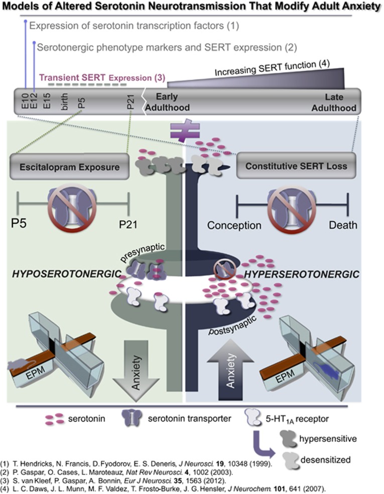
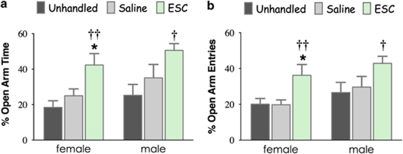
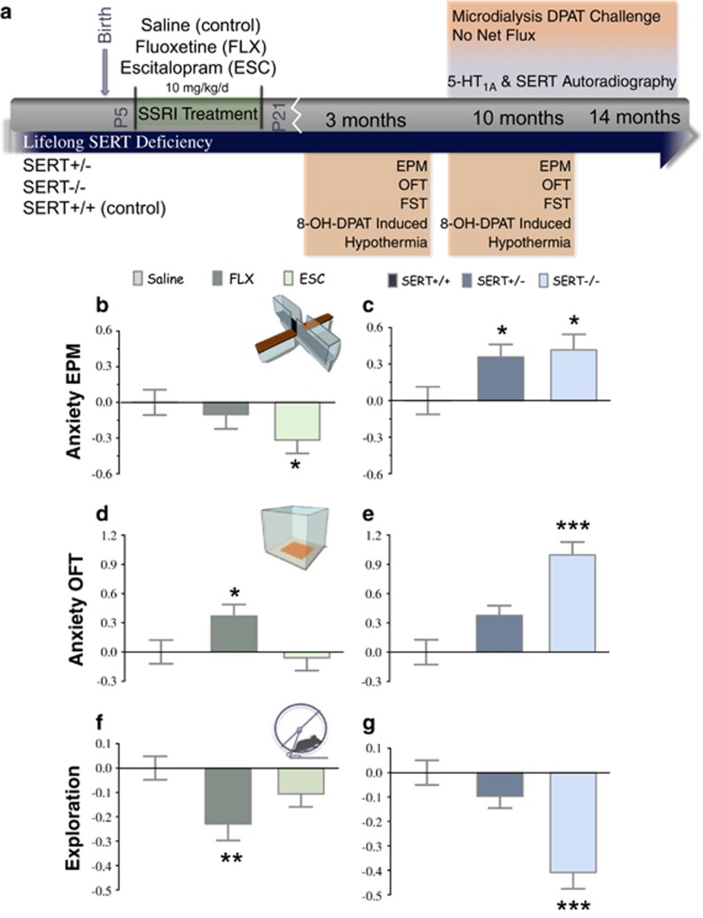
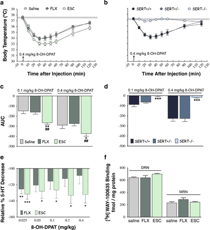
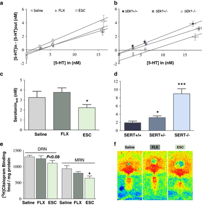
Large numbers of women undergo antidepressant treatment during pregnancy; however, long-term consequences for their offspring remain largely unknown. Rodents exposed to serotonin transporter (SERT)-inhibiting antidepressants during development show changes in adult emotion-like behavior. These changes have been equated with behavioral alterations arising from genetic reductions in SERT. Both models are highly relevant to humans yet they vary in their time frames of SERT disruption. We find that anxiety-related behavior and, importantly, underlying serotonin neurotransmission diverge between the two models. In mice, constitutive loss of SERT causes life-long increases in anxiety-related behavior and hyperserotonemia. Conversely, early exposure to the antidepressant escitalopram (ESC; Lexapro) results in decreased anxiety-related behavior beginning in adolescence, which is associated with adult serotonin system hypofunction in the ventral hippocampus. Adult behavioral changes resulting from early fluoxetine (Prozac) exposure were different from those of ESC and, although somewhat similar to SERT deficiency, were not associated with changes in hippocampal serotonin transmission in late adulthood. These findings reveal dissimilarities in adult behavior and neurotransmission arising from developmental exposure to different widely prescribed antidepressants that are not recapitulated by genetic SERT insufficiency. Moreover, they support a pivotal role for serotonergic modulation of anxiety-related behavior.


Figures

Figure 1
Figure 1
Different time frames of serotonin transporter (SERT) disruption and effects on serotonin transmission and anxiety-related behavior. Synthesis of brain serotonin begins during early embryonic development in mice (embryonic (E) day 12.5) and is preceded by the expression of transcription factors, ie, Pet-1 ETS, Lmx1b, and Gata3, necessary for serotonergic phenotype determination (1 and 2). Serotonin transporter expression is observed as early as E12.5 in brain stem serotonin neurons and also occurs briefly in nonserotonergic neurons (∼E13 through postnatal (P) day 21), predominately in thalamic glutamatergic neurons projecting to cortical sensory processing areas (3). Serotonin transporter expression and function continue to increase throughout adulthood (4). We hypothesized that SERT inhibition limited to early postnatal development (P5-P21) via exposure to selective serotonin reuptake inhibitors (SSRIs) in mice would produce changes in serotonin system function and behavior different from those arising from constitutive SERT deficiency, which alters SERT function throughout life. We discovered that postnatal administration of the antidepressant escitalopram (ESC) but not fluoxetine results in a hyposerotonergic state during adulthood associated with reduced anxiety-related behavior in the elevated plus maze (EPM). This directly contrasts with the hyperserotonergic state (increased extracellular serotonin levels) and increased anxiety-like behavior in mice with lifelong reductions in Sert gene expression. Hyposerotonemia is accompanied by 5-HT1A autoreceptor hypersensitivity in mice exposed to postnatal ESC, while genetic SERT deficiency causes desensitization of 5-HT1A autoreceptors.
Figure 2
Figure 2
Anxiety-related behavior in adolescent mice after early postnatal exposure to escitalopram. Administration of escitalopram to female and male mice during P5–P21 increased (a) the percentage of open arm time and (b) the percentage of open arm entries in the elevated plus maze compared with saline-treated and unhandled mice during periadolescence (P50). *P<0.05 vs unhandled control; P<0.05 and ††P<0.01 vs saline-treated control.
Figure 3
Figure 3
Anxiety-related and exploratory behavior in postnatal SSRI-exposed vs SERT-deficient mice. (a) The timeline shows the treatment paradigm for postnatal SSRI administration and the timing of experimental testing for adult SERT-deficient and SSRI-exposed mice. For global behavior analysis, standard score normalization was applied with respect to age and sex for each variable to combine data with different dynamic ranges and directions of change (Guilloux et al, 2011). Standard scores were weighted by coefficients proportional to their loadings in the factor analysis (Supplementary Table S3). (b and c) Latency to first open arm entry, open arm time, the percentage of open arm entries, and head dips in the elevated plus maze loaded on a single factor (‘Anxiety EPM'). (d and e) The open field anxiety-like parameters, center time, and ratio of center to total distance traveled loaded on a separate factor (‘Anxiety OFT'). (f and g) Exploration (factor 3) was determined by total arm entries and rears in the elevated plus maze and total distance traveled in the open field. Group sizes are listed in Supplementary Table S1. *P<0.05, **P<0.01, and ***P<0.001 vs postnatal saline-treated or SERT+/+ mice.
Figure 4
Figure 4
Serotonin-1A autoreceptor function and expression in postnatal SSRI-exposed vs SERT-deficient mice in late adulthood. (a and b) Time courses show changes in body temperature associated with administration of 0.4 mg/kg 8-OH-DPAT to 10-month-old male postnatal SSRI-exposed and SERT-deficient mice. Gray-shaded horizontal bars represent mean body temperatures±SEMs for saline challenge across postnatal treatment groups or genotypes. (c and d) Potentiated hypothermia after 0.1 mg/kg or 0.4 mg/kg 8-OH-DPAT in 10-month-old male mice exposed to postnatal ESC is indicated by increased areas under the curve. Conversely, a complete absence of thermal response after both doses of 8-OH-DPAT was observed in 10-month-old male SERT−/− mice. (e) Percentage of decreases in extracellular serotonin levels in the ventral hippocampus of freely moving 10–14-month-old postnatal SSRI-exposed mice in response to 8-OH-DPAT challenge relative to decreases occurring in postnatal saline-treated mice. (f) Specific binding of [3H]WAY-100635 in the dorsal raphe nucleus (DRN) or medial raphe nucleus (MRN) with respect to postnatal SSRI administration. For temperature and microdialysis data, N=11, 16, and 17 for postnatal saline-, FLX-, and ESC-treated mice, respectively, and N=13, 19, and 12 for SERT+/+, SERT+/−, and SERT−/− mice, respectively. For autoradiography, N=3–5 mice/group. For hypothermia data, *P<0.05 and **P<0.01 vs saline-treated mice, ##P<0.01 vs FLX-treated mice, and ***P<0.001 vs SERT+/+ mice. For microdialysis data, *P<0.05, **P<0.01, and ***P<0.001 vs postnatal saline-treated mice administered the same dose of 8-OH-DPAT.
Figure 5
Figure 5
Extracellular serotonin concentrations and serotonin transporter expression. (a and b) Linear regression (no-net-flux) analysis was performed on different concentrations of serotonin perfused into microdialysis probes (Cin) minus dialysate serotonin concentrations (Cout) (y axis) vs Cin (x axis). Intercepts (x axis) were used to estimate extracellular serotonin levels in ventral hippocampus corrected for in vivo extraction fraction. Error bars are too small to be pictured in some cases. (c and d) Extracellular serotonin was reduced in postnatal ESC-exposed mice compared with postnatal saline-exposed mice at 10–14 months of age. By contrast, extracellular serotonin was elevated in SERT+/− and SERT−/− mice vs SERT+/+ mice. Extracellular serotonin levels were 3.3±0.6 nM, 3.8±0.5 nM, and 2.2±0.3 nM in saline- (N=15), FLX- (N=17), and ESC-treated (N=19) mice, respectively. Mean extracellular serotonin levels for SERT+/+ (N=14), SERT+/− (N=19), and SERT−/− (N=15) mice were 1.9±0.4 nM, 3.2±0.4 nM, and 9.0±1.2 nM, respectively. (e and f) Total SERT binding was reduced in the median raphe nucleus (MRN) in postnatal ESC-treated mice (N=4–5). Representative autoradiograms are shown for each treatment group. *P<0.05 vs postnatal saline-treated mice or SERT+/+ mice, and **P<0.01 and ***P<0.001 vs SERT+/+ mice.

References

    1. Altieri S, Singh Y, Sibille E, Andrews A.2012Serotonergic pathways in depressionIn: Lopez-Munoz F, Alamo C (eds). Neurobiology of Depression CRC Press, Taylor & Francis Group: Boca Raton, FL, USA; 143–170.
    1. Altieri SC, Garcia-Garcia AL, Leonardo ED, Andrews AM. Rethinking 5-HT(1A) receptors: emerging modes of inhibitory feedback of relevance to emotion-related behavior. ACS Chem Neurosci. 2013;4:72–83. - PMC - PubMed
    1. Ansorge MS, Morelli E, Gingrich JA. Inhibition of serotonin but not norepinephrine transport during development produces delayed, persistent perturbations of emotional behaviors in mice. J Neurosci. 2008;28:199–207. - PMC - PubMed
    1. Ansorge MS, Zhou M, Lira A, Hen R, Gingrich JA. Early-life blockade of the 5-HT transporter alters emotional behavior in adult mice. Science. 2004;306:879–881. - PubMed
    1. Beikmann BS, Tomlinson ID, Rosenthal SJ, Andrews AM. Serotonin uptake is largely mediated by platelets versus lymphocytes in peripheral blood cells. ACS Chem Neurosci. 2013;4:161–170. - PMC - PubMed

Publication types

MeSH terms