. 2015 Feb 15;308(4):G325-34.
doi: 10.1152/ajpgi.00429.2013. Epub 2014 Dec 18.

Role of Fn14 in acute alcoholic steatohepatitis in mice

Affiliations

Role of Fn14 in acute alcoholic steatohepatitis in mice

Gamze Karaca et al. Am J Physiol Gastrointest Liver Physiol. .

Abstract

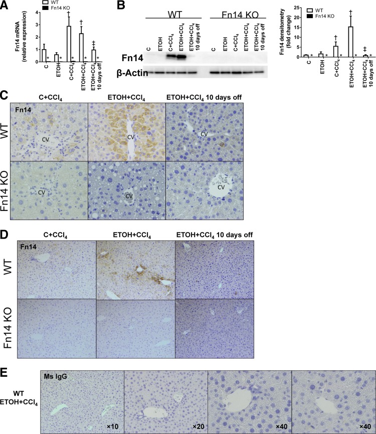
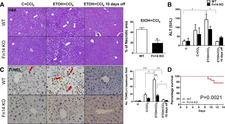
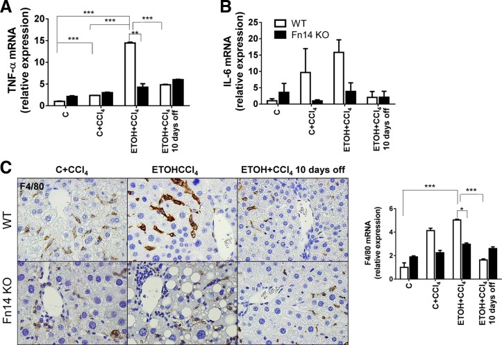
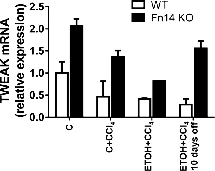
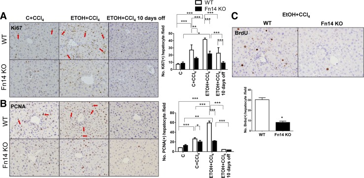
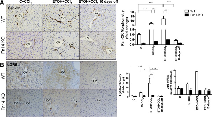
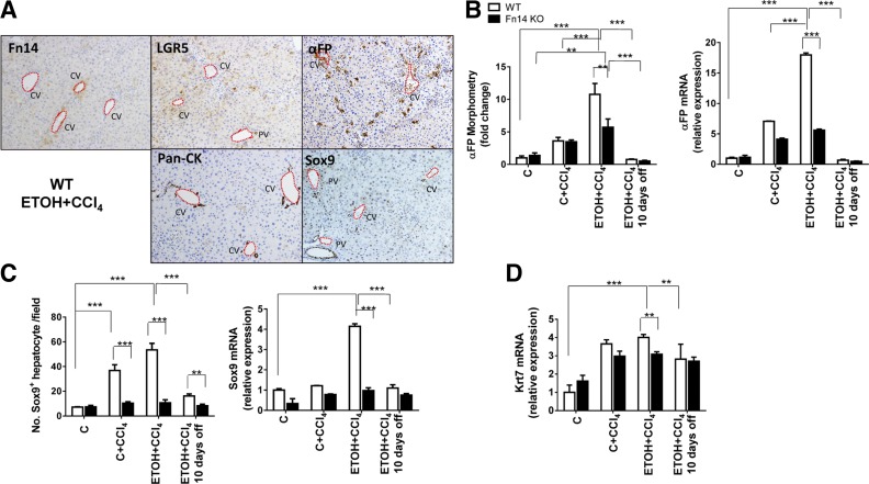
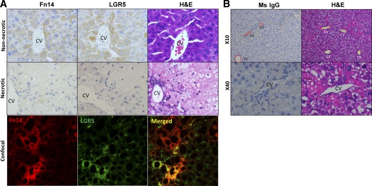
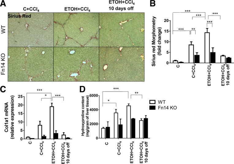
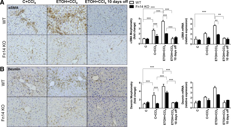
TNF-like weak inducer of apoptosis (TWEAK) is a growth factor for bipotent liver progenitors that express its receptor, fibroblast growth factor-inducible 14 (Fn14), a TNF receptor superfamily member. Accumulation of Fn14(+) progenitors occurs in severe acute alcoholic steatohepatitis (ASH) and correlates with acute mortality. In patients with severe ASH, inhibition of TNF-α increases acute mortality. The aim of this study was to determine whether deletion of Fn14 improves the outcome of liver injury in alcohol-consuming mice. Wild-type (WT) and Fn14 knockout (KO) mice were fed control high-fat Lieber deCarli diet or high-fat Lieber deCarli diet with 2% alcohol (ETOH) and injected intraperitoneally with CCl₄ for 2 wk to induce liver injury. Mice were euthanized 3 or 10 days after CCl₄ treatment. Survival was assessed. Liver tissues were analyzed for cell death, inflammation, proliferation, progenitor accumulation, and fibrosis by quantitative RT-PCR, immunoblot, hydroxyproline content, and quantitative immunohistochemistry. During liver injury, Fn14 expression, apoptosis, inflammation, hepatocyte replication, progenitor and myofibroblast accumulation, and fibrosis increased in WT mice fed either diet. Mice fed either diet expressed similar TWEAK/Fn14 levels, but ETOH-fed mice had higher TNF-α expression. The ETOH-fed group developed more apoptosis, inflammation, fibrosis, and regenerative responses. Fn14 deletion did not reduce hepatic TNF-α expression but improved all injury parameters in mice fed the control diet. In ETOH-fed mice, Fn14 deletion inhibited TNF-α induction and increased acute mortality, despite improvement in liver injury. Fn14 mediates wound-healing responses that are necessary to survive acute liver injury during alcohol exposure.

Keywords: alcohol; liver fibrosis; liver injury; liver progenitors.


Figures

Fig. 1.
Fig. 1.
Subacute liver injury triggers hepatic accumulation of fibroblast growth factor-inducible 14 (Fn14)-positive (Fn14+) cells. Healthy adult wild-type (WT) mice fed control, high-fat Lieber deCarli diet (C) or high-fat Lieber deCarli diet with 2% alcohol (ETOH) were treated with CCl4 for 2 wk and euthanized at the time points described in materials and methods. Expression of hepatic Fn14 was evaluated by quantitative RT-PCR analysis (normalized to the housekeeping gene S9; A), representative Western blot analysis for Fn14 (normalized to β-actin loading control; B, left), and quantification of Western blot analysis by densitometry (B, right) and representative immunohistochemistry (C; magnification ×40). To ensure reagent specificity, similar analysis was performed in mice that were genetically deficient in Fn14 [Fn14 knockout (KO)]. CV, central vein; PV, portal vein. D: representative immunohistochemistry of Fn14 in liver sections from WT and Fn14 KO mice at lower magnification (original magnification ×10). E: representative images from immunohistochemistry of a mouse IgG (Ms IgG) control on liver sections from WT mice fed ETOH diet and treated with CCl4. *P < 0.05 vs. WT. †P < 0.05 vs. C. ‡P < 0.05 vs. ETOH + CCl4.
Fig. 2.
Fig. 2.
Deletion of Fn14 increases mortality, despite improving liver injury. A–D: liver histology (A), serum alanine aminotransferase (ALT) levels (B), terminal deoxynucleotidyl transferase dUTP nick end label (TUNEL) staining [red arrows indicate TUNEL+ cells; C], and survival (D) in WT and Fn14 KO mice fed control or ETOH diet and treated with intraperitoneal injections of CCl4 for 2 wk. A: hematoxylin-eosin stained sections of livers from WT mice fed ETOH diet and treated with CCl4 and similarly treated Fn14 KO mice. Percent necrosis was quantified from 50 randomly chosen fields per section in ETOH + CCl4 groups. Comparisons for ALT and TUNEL+ hepatocytes per field were caculated by 2-way ANOVA followed by Tukey's multiple-comparison post hoc test: *P < 0.05, ***P < 0.001. Significance for survival curve was calculated using log-rank test.
Fig. 3.
Fig. 3.
Deletion of Fn14 reduces proinflammatory cytokine expression. A and B: whole liver mRNA expression of the inflammatory cytokines TNF-α and IL-6 in WT and Fn14 KO mice. Results were normalized to the housekeeping gene S9. C: representative immunohistochemistry (original magnification ×40) and mRNA expression for the macrophage marker F4/80. Results are expressed as fold change compared with control. Comparisons for TNF-α and F4/80 were calculated by 2-way ANOVA followed by Tukey's multiple-comparison post hoc test: *P < 0.05, **P < 0.01, ***P < 0.001. One-way ANOVA showed that IL-6 levels differ significantly across exposure in WT (P = 0.04), but not Fn14 KO, mice.
Fig. 4.
Fig. 4.
ETOH feeding + CCl4 injection does not induce hepatic expression of the Fn14 ligand TNF-like weak inducer of apoptosis (TWEAK). Expression of hepatic TWEAK was evaluated by quantitative RT-PCR analysis in livers from healthy adult WT mice fed control or ETOH diet, treated with CCl4 for 2 wk, and euthanized at the time points described in materials and methods. Results were normalized to expression of the housekeeping gene S9. One-way ANOVA showed that TWEAK levels did not differ significantly across exposure in WT or Fn14 KO mice.
Fig. 5.
Fig. 5.
Deletion of Fn14 impairs hepatocyte proliferation. Liver sections from WT and Fn14 KO mice were stained for Ki67 (A), proliferating cell nuclear antigen (PCNA, B), and bromodeoxyuridine (BrdU; C). Original magnification ×10. Red arrows indicate Ki67+ or PCNA+ hepatocytes. Positive hepatocytes were quantified in 10 randomly selected fields per section per mouse. Comparisons for Ki67+ and PCNA+ hepatocytes per field were calculated by 2-way ANOVA followed by Tukey's multiple-comparison post hoc test: *P < 0.05, **P < 0.01, ***P < 0.001.
Fig. 6.
Fig. 6.
Deletion of Fn14 inhibits liver progenitor accumulation. Liver sections from WT and Fn14 KO mice were stained for pan-cytokeratin (pan-CK; A) and LGR5 (B). Original magnification ×20. CV, central vein; PV, portal vein. Morphometric data and LGR5 mRNA expression in the respective groups are shown. Results are expressed as fold change compared with control. Results were normalized to the housekeeping gene S9. All comparisons were calculated by 2-way ANOVA followed by Tukey's multiple-comparison post hoc test: *P < 0.05, ***P < 0.001.
Fig. 7.
Fig. 7.
Deletion of Fn14 impairs liver progenitor expansion. A: representative immunohistochemistry of Fn14, LGR5, α-fetoprotein (αFP), pan-CK, and Sox9 in liver sections from WT mice fed ETOH diet and treated with CCl4 for 2 wk. Original magnification ×10. CV, central vein; PV, portal vein. B–D: morphometry data and mRNA expression for αFP (B) and Sox9 (C) and mRNA expression for keratin 7 (Krt7; D) for livers from WT and Fn14 KO mice. Results are expressed as fold change compared with control. Results were normalized to the housekeeping gene S9. All comparisons were calculated by 2-way ANOVA followed by Tukey's multiple-comparison post hoc test: **P < 0.01, ***P < 0.001.
Fig. 8.
Fig. 8.
Liver Fn14+ cells coexpress LGR5. A: serial section staining for Fn14, LGR5, and hematoxylin-eosin (H&E) from nonnecrotic and necrotic regions in WT mice after injury induced by CCl4 and ETOH (top and middle) and confocal image of Fn14 and LGR5 in injured liver (bottom). Original magnification, ×40. B: representative images from immunohistochemistry of a mouse IgG (Ms IgG) control for Fn14 and hematoxylin-eosin on necrotic area in liver sections from WT mice fed ETOH diet and treated with CCl4. CV, central vein; PV, portal vein.
Fig. 9.
Fig. 9.
Deletion of Fn14 reduces liver fibrosis. Liver sections from WT and Fn14 KO mice were stained with Sirius red to demonstrate fibrosis (A; original magnification ×10), with morphometry displayed as means ± SE (B). C: quantitative RT-PCR analysis of collagen 1α1 (Col 1α1) mRNA levels, with results normalized to the housekeeping gene S9. D: hepatic hydroxyproline content in the respective groups. Results are expressed as fold change compared with control. All comparisons were calculated by 2-way ANOVA followed by Tukey's multiple-comparison post hoc test: *P < 0.05, **P < 0.01, ***P < 0.001.
Fig. 10.
Fig. 10.
Deletion of Fn14 reduces myofibroblast accumulation. A and B: representative immunohistochemistry (original magnification × 20), morphometry, and mRNA expression for αSMA and desmin. Results are expressed as fold change compared with control. All comparisons were calculated by 2-way ANOVA followed by Tukey's multiple-comparison post hoc test: **P < 0.01, ***P < 0.001.

References

    1. Affo S, Dominguez M, Lozano JJ, Sancho-Bru P, Rodrigo-Torres D, Morales-Ibanez O, Moreno M, Millan C, Loaeza-del-Castillo A, Altamirano J, Garcia-Pagan JC, Arroyo V, Gines P, Caballeria J, Schwabe RF, Bataller R. Transcriptome analysis identifies TNF superfamily receptors as potential therapeutic targets in alcoholic hepatitis. Gut 62: 452–460, 2013. - PMC - PubMed
    1. An L, Wang X, Cederbaum AI. Cytokines in alcoholic liver disease. Arch Toxicol 86: 1337–1348, 2012. - PubMed
    1. Bird TG, Lu WY, Boulter L, Gordon-Keylock S, Ridgway RA, Williams MJ, Taube J, Thomas JA, Wojtacha D, Gambardella A, Sansom OJ, Iredale JP, Forbes SJ. Bone marrow injection stimulates hepatic ductular reactions in the absence of injury via macrophage-mediated TWEAK signaling. Proc Natl Acad Sci USA 110: 6542–6547, 2013. - PMC - PubMed
    1. Boulter L, Govaere O, Bird TG, Radulescu S, Ramachandran P, Pellicoro A, Ridgway RA, Seo SS, Spee B, Van Rooijen N, Sansom OJ, Iredale JP, Lowell S, Roskams T, Forbes SJ. Macrophage-derived Wnt opposes Notch signaling to specify hepatic progenitor cell fate in chronic liver disease. Nat Med 18: 572–579, 2012. - PMC - PubMed
    1. Diehl AM, Yin M, Fleckenstein J, Yang SQ, Lin HZ, Brenner DA, Westwick J, Bagby G, Nelson S. Tumor necrosis factor-α induces c-jun during the regenerative response to liver injury. Am J Physiol Gastrointest Liver Physiol 267: G552–G561, 1994. - PubMed

Publication types

MeSH terms