Review
. 2015 Mar;24(3):298-309.
doi: 10.1002/pro.2626. Epub 2015 Jan 13.

The structural basis for specificity in lipoxygenase catalysis

Affiliations
Review

The structural basis for specificity in lipoxygenase catalysis

Marcia E Newcomer et al. Protein Sci. 2015 Mar.

Abstract

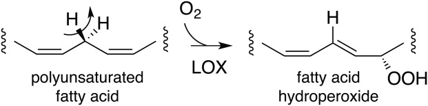
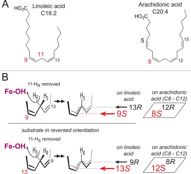
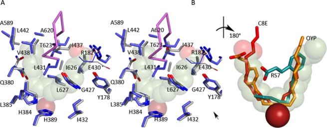
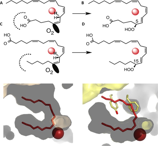
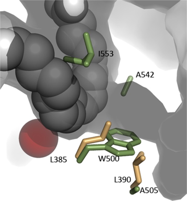
Many intriguing facets of lipoxygenase (LOX) catalysis are open to a detailed structural analysis. Polyunsaturated fatty acids with two to six double bonds are oxygenated precisely on a particular carbon, typically forming a single chiral fatty acid hydroperoxide product. Molecular oxygen is not bound or liganded during catalysis, yet it is directed precisely to one position and one stereo configuration on the reacting fatty acid. The transformations proceed upon exposure of substrate to enzyme in the presence of O2 (RH + O2 → ROOH), so it has proved challenging to capture the precise mode of substrate binding in the LOX active site. Beginning with crystal structures with bound inhibitors or surrogate substrates, and most recently arachidonic acid bound under anaerobic conditions, a picture is consolidating of catalysis in a U-shaped fatty acid binding channel in which individual LOX enzymes use distinct amino acids to control the head-to-tail orientation of the fatty acid and register of the selected pentadiene opposite the non-heme iron, suitably positioned for the initial stereoselective hydrogen abstraction and subsequent reaction with O2 . Drawing on the crystal structures available currently, this review features the roles of the N-terminal β-barrel (C2-like, or PLAT domain) in substrate acquisition and sensitivity to cellular calcium, and the α-helical catalytic domain in fatty acid binding and reactions with O2 that produce hydroperoxide products with regio and stereospecificity. LOX structures combine to explain how similar enzymes with conserved catalytic machinery differ in product, but not substrate, specificities.

Keywords: arachidonic acid; hydroperoxide oxygenation; linoleic acid; lipoxygenase; oxylipins; polyunsaturated fatty acids.


Figures

Scheme 1
Scheme 1
Scheme Lipoxygenase catalysis.
Figure 1
Figure 1
Figure The basis of LOX regio- and stereospecificity. (A)The structures of linoleic acid (18:2) and arachidonic acid (20:4). (B) A perspective view on LOX catalysis showing the reacting pentadiene. Top left: The catalytic iron (Fe-OH) abstracts the pro-R hydrogen from C11 and O2 is added antarafacially in the 9S position (in red, in the foreground). Lower left: with linoleic acid in a reversed head-to-tail orientation, the identical stereochemical relationships involve pro-S hydrogen abstraction at C11 and oxygenation in the 13S position (in red, in the foreground). Formation of the corresponding 9R or 13R configuration products entails antarafacial reaction of O2 at the opposite end of the pentadiene. In boxes on the right side: an example of the corresponding positional specificities of arachidonic acid, illustrating the positions around the 8–12 pentadiene on arachidonic acid.
Figure 2
Figure 2
Figure The common core of LOXs is shared by plant, animal and bacterial enzymes. (A) The core domain is a large bundle of helices that houses the catalytic iron. A distinct insertion that contributes to a helix that forms an arch over the active site is colored in bright pink. (B) and (C) The animal (flesh) and plant (green) enzymes have amino terminal domain PLAT domains that may harbor Ca2+ binding sites. (D) The bacterial LOX lacks the PLAT domain, but has additional helices. An interactive view is available in the electronic version of the article.
Figure 3
Figure 3
Figure Four helices form the heart of the common core structure (depicted in 8R-LOX). Amino acids from these helices contribute the major part of the side chains that form the iron binding site and substrate binding cavity. (A) The location of these four helices in the LOX overall structure. (B) The amino acids that form the arachidonic acid binding site in 8R, colored accordingly. (C) and (D) Details of the helical core with 90° rotation so that the cavity entrance can be visualized. An interactive view is available in the electronic version of the article.
Figure 4
Figure 4
Figure The iron coordination sphere. (A) Superposition of the iron ligands from representative plant (1YGE) animal (4QWT) and bacterial (4G32) structures. (B) The C-terminal region of the enzymes; a long loop that follows the terminal α-helix makes its way to the catalytic iron so that the carboxyl can fill the coordination sphere. (C). A portion of the electron density map for 8R-LOX (3FG1), centered around the C-terminal Ile. Note the presence of a water-filled cavity that accommodates the terminal Ile.
Figure 5
Figure 5
Figure The placement of α2 varies among LOXs. The “arched” helix and helix α2 in the plant (green, 1YGE) animal (flesh, 4QWT) and bacterial enzymes (light blue, 4G32) are superimposed. The “broken” α2 (brown, 3O8Y) of 5-LOX is included as well for reference. Note that while the arched helices from the diverse structure superpose, α2 varies in its placement.
Figure 6
Figure 6
Figure The active site of animal LOXs can be “corked.” (A) The corking amino acids of 5-LOX (pink) and 11R-LOX (yellow) plug what would otherwise be an open U-shaped cavity (15-LOX-2, flesh). (B) The corking amino acids are conserved in “uncorked” LOXs (blue, 12-LOX, green, 15-LOX-2), however since α2 is not “broken” the side chains are distal and cannot seal the active site entrance. The image has been rotated relative to (A) for clarity. (C) In the inhibited structure of 12-LOX an aromatic ring of the inhibitor (gold, OYP) occupies the position of the “cork.”
Figure 7
Figure 7
Figure Ca2+ binding appears to promote structural changes in the loops of the PLAT domain. (A) Superposition of cartoon renderings of 8R-LOX (light blue) and 15-LOX-2 structures (flesh), both in the presence of Ca2+. Three Ca2+ (spheres) sites are found in the former, and two in the latter. (B) Superposition of 8R-LOX in the presence of Ca2+ and 11-R-LOX in its absence. Despite a higher sequence identity in this pair of LOX, the loop conformations are divergent, presumably a reflection of differences in the Ca2+-bound- and Ca2+-free states.
Figure 8
Figure 8
Figure (A) The substrate adopts a horseshoe shape in the U-shaped channel (stereo). The side chains of highly conserved amino acids line the base of the active site, along with Gly-427. Glu-430, part of an inter-helical charge cluster that includes the substrate carboxylate, is shown in line rendering. The Fe2+ (transparent rust sphere) is positioned behind the substrate. (B) Detail of the superposition of inhibitors (rotated ∼180° with respect to (A)) observed in 15-LOX-2 (red) 12-LOX (gold C, red O) and 15-LOX-1 (teal C, red O). The 15-LOX-2 and 12-LOX inhibitors conform to the arachidonic acid placement, while the 15-LOX-1 inhibitor overlaps partially. The Fe2+, solid rust sphere, is in front of the substrate in this view. An interactive view is available in the electronic version of the article.
Figure 9
Figure 9
Figure Inverse entry of substrate is consistent with cavity depth in 15-LOX-2 and 5-LOX. (A) A schematic depicting how the products 15S-HPETE and 5S-HPETE can be explained by substrate entry in 15-LOX-2 as “tail-first” and that in 5-LOX as “head-first.” (B) The cavity depths in 15-LOX-2 (flesh) and 5-LOX (yellow) are consistent with this interpretation. The innermost part of the 5-LOX cavity should accommodate a carboxyl, rather than hydrocarbon.
Figure 10
Figure 10
Figure A possible O2 access channel intersects with the U-shaped cavity on the side opposite the catalytic iron in 8R-LOX. Amino acids which have been shown to control O2 access in other LOXs are in included. Soybean LOX1 side chains (green) are Ile-553, Ala-542 (the Gly/Ala switch), Trp-500 and Ala-505. Leu-385 and Leu-390 are the animal counterparts of the Trp-Ala pair in plants.

References

    1. Ivanov I, Heydeck D, Hofheinz K, Roffeis J, O'Donnell VB, Kuhn H, Walther M. Molecular enzymology of lipoxygenases. Arch Biochem Biophys. 2010;503:161–174. - PubMed
    1. Samuelsson B, Dahlen SE, Lindgren JA, Rouzer CA, Serhan CN. Leukotrienes and lipoxins: structures, biosynthesis, and biological effects. Science. 1987;237:1171–1176. - PubMed
    1. Andreou A, Feussner I. Lipoxygenases—structure and reaction mechanism. Phytochemistry. 2009;70:1504–1510. - PubMed
    1. Zheng Y, Yin H, Boeglin WE, Elias PM, Crumrine D, Beier DR, Brash AR. Lipoxygenases mediate the effect of essential fatty acid in skin barrier formation: a proposed role in releasing omega-hydroxyceramide for construction of the corneocyte lipid envelope. J Biol Chem. 2011;286:24046–24056. - PMC - PubMed
    1. Meyer D, Herrfurth C, Brodhun F, Feussner I. Degradation of lipoxygenase-derived oxylipins by glyoxysomes from sunflower and cucumber cotyledons. BMC Plant Biol. 2013;13:177. - PMC - PubMed

Publication types