Comparative Study
. 2014 Dec 23;64(24):2589-2600.
doi: 10.1016/j.jacc.2014.09.059.

Clinical phenotype and outcome of hypertrophic cardiomyopathy associated with thin-filament gene mutations

Affiliations
Comparative Study

Clinical phenotype and outcome of hypertrophic cardiomyopathy associated with thin-filament gene mutations

Raffaele Coppini et al. J Am Coll Cardiol. .

Abstract

Background: Mild hypertrophy but increased arrhythmic risk characterizes the stereotypic phenotype proposed for hypertrophic cardiomyopathy (HCM) caused by thin-filament mutations. However, whether such clinical profile is different from more prevalent thick-filament-associated disease is unresolved.

Objectives: This study aimed to assess clinical features and outcomes in a large cohort of patients with HCM associated with thin-filament mutations compared with thick-filament HCM.

Methods: Adult HCM patients (age >18 years), 80 with thin-filament and 150 with thick-filament mutations, were followed for an average of 4.5 years.

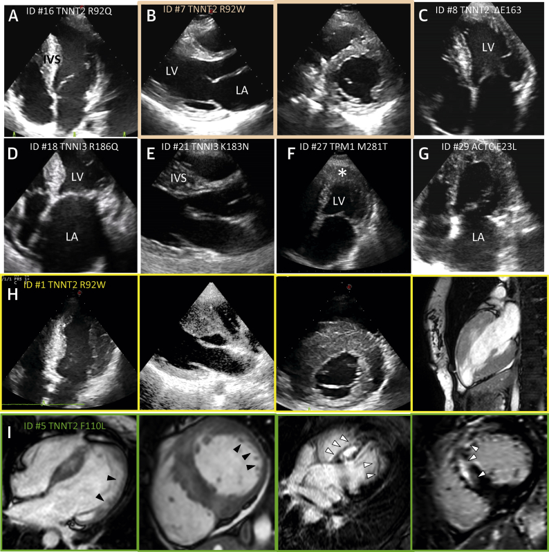
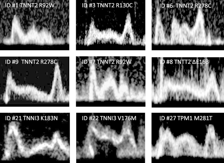
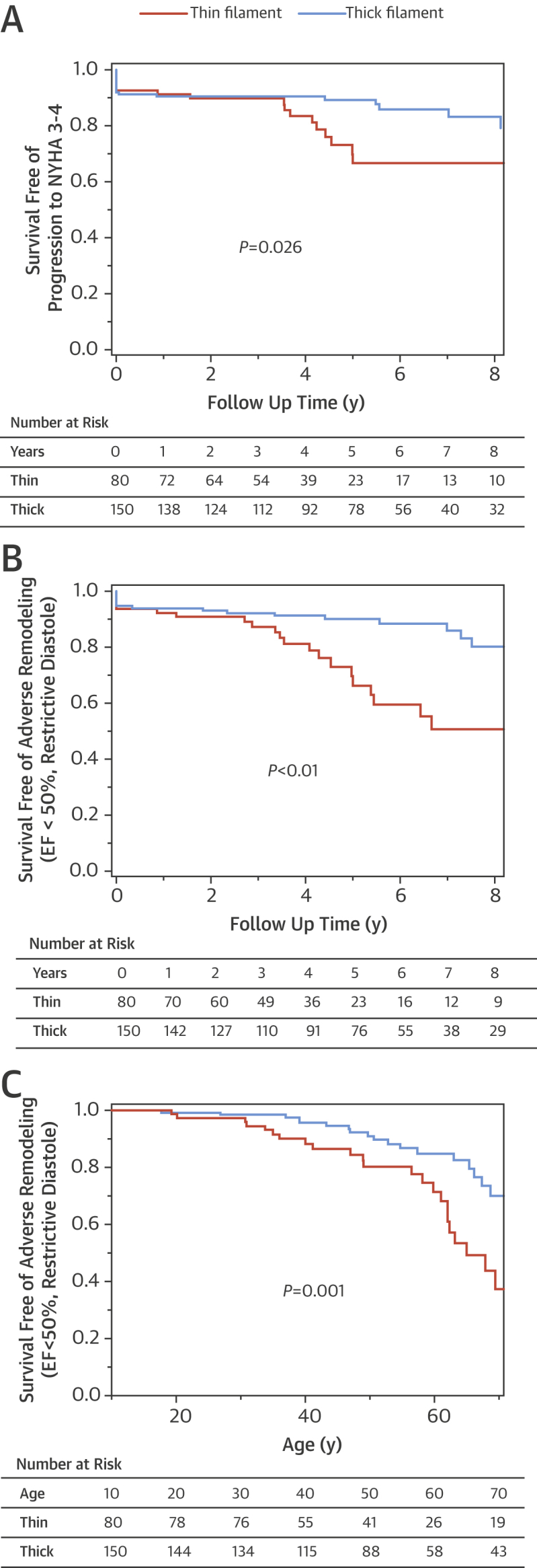
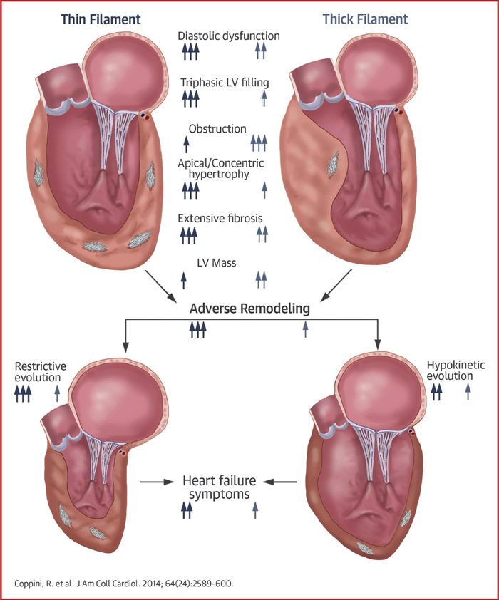
Results: Compared with thick-filament HCM, patients with thin-filament mutations showed: 1) milder and atypically distributed left ventricular (LV) hypertrophy (maximal wall thickness 18 ± 5 mm vs. 24 ± 6 mm; p < 0.001) and less prevalent outflow tract obstruction (19% vs. 34%; p = 0.015); 2) higher rate of progression to New York Heart Association functional class III or IV (15% vs. 5%; p = 0.013); 3) higher prevalence of systolic dysfunction or restrictive LV filling at last evaluation (20% vs. 9%; p = 0.038); 4) 2.4-fold increase in prevalence of triphasic LV filling pattern (26% vs. 11%; p = 0.002); and 5) similar rates of malignant ventricular arrhythmias and sudden cardiac death (p = 0.593).

Conclusions: In adult HCM patients, thin-filament mutations are associated with increased likelihood of advanced LV dysfunction and heart failure compared with thick-filament disease, whereas arrhythmic risk in both subsets is comparable. Triphasic LV filling is particularly common in thin-filament HCM, reflecting profound diastolic dysfunction.

Keywords: diastolic function; end-stage; genotype to phenotype correlation; triphasic filling; troponin.


Figures

Figure 1
Figure 1
The Cardiac Sarcomere Thin Filament Schematic representation of the thin filament and its key molecular components (colored) in relation to thick-filament proteins (gray). Thin-filament proteins with disease-causing mutations found in the thin-filament cohort of this study are circled in red.
Figure 2
Figure 2
Genetic Basis of Thin-Filament HCM Exons and structural and functional domains of cardiac troponin T (cTnT) and I (cTnI), tropomyosin (Tm), and actin, showing mutation sites identified in the study cohort. Black, certainly pathogenic mutations (see Methods); slate-grey, mutations likely to be pathogenic. For mutations carried by more than 1 patient, the total number of carriers is indicated. HCM = hypertrophic cardiomyopathy.
Figure 3
Figure 3
Phenotypic Variability in Thin-Filament–Associated HCM Echocardiographic and cardiomagnetic resonance (CMR) images showing variable extent and distribution of left ventricular hypertrophy (LVH) in patients from the thin-filament study cohort (patient’s ID number and mutation are indicated). (A) Moderate LVH associated with TNNT2-R92Q, with classic localization at the interventricular septum (IVS). (B) Mild midseptal hypertrophy and marked left atrial (LA) dilation associated with TNNT2-ΔE163. (C) Mild concentric LVH associated with TNNT2-R92W. (D) Moderate midseptal LVH with small LV cavity size and severe LA dilation (restrictive evolution) associated with TNNI3-R186Q. (E) Classic asymmetric septal LVH associated with TNNI3-K183M. (F) Apical LVH associated with TPM-M281T; indicates LV apex. (G) Apical LVH with severe LA dilation associated with ACTC-F23L. (H) Markedly asymmetric LVH involving the anterior and posterior septum and anterior wall associated with TNNT2-R92W. (I) CMR images from a patient carrying the TNNT2-F110L mutation. (From left) Four-chamber and midventricular short-axis views showing extensive regions of noncompaction (black arrowheads) in the LV apical and free wall regions; 4-chamber and short-axis views showing large areas of late gadolinium enhancement (white arrowheads) in the anterior free wall and in the septum, respectively. HCM = hypertrophic cardiomyopathy.
Figure 4
Figure 4
Triphasic LV Filling Pattern Representative transmitral blood flow velocity patterns assessed by pulsed-wave Doppler echocardiography in 9 patients with thin-filament–associated hypertrophic cardiomyopathy (patient ID numbers and mutations are shown). Mid-diastolic flow velocity (L-wave) is clearly visible (*). An L wave’s presence is independent of the overall diastolic pattern, with E/A ratios that can be <1 (i.e., delayed relaxation) or >1 (i.e., pseudonormalized). LV = left ventricular.
Figure 5
Figure 5
Clinical and Instrumental Outcomes in Thick- Versus Thin-Filament HCM (A) Kaplan-Mayer curve illustrating survival free of progression to severe heart failure (New York Health Association [NYHA] functional classes III/IV). (B) Survival free of adverse left ventricular (LV) remodeling and dysfunction during follow-up, defined as progression to LV ejection fraction (EF) <50% or toward restrictive LV filling pattern. (C) Lifelong likelihood of advanced LV dysfunction (defined as in B) in relation to genetic status. The probability values are calculated with the log-rank test comparing thin-filament versus thick-filament survival curves. HCM = hypertrophic cardiomyopathy; NYHA = New York Heart Association.
Figure 6
Figure 6
Evidence of Disease Progression in Thin-filament HCM (A) Echocardiographic images from patient ID #5 carrying the TNNT2-F110L mutation. Top: Echocardiographic evaluation at age 16 years. (Left) Parasternal short-axis view showing severe and diffuse anteroseptal LV hypertrophy. (Right) Parasternal long-axis view showing turbulent flow in the LV outflow tract (arrow), caused by severe dynamic obstruction. (Bottom) Similar views from the same patient at age 37 years, showing marked anteroseptal wall thinning and absence of obstruction and increased left atrial size. (B) CMR images from patient ID #32 carrying the TNNT2-R92W mutation. (Top) Cardiac magnetic resonance at age 21 years. Short-axis and 3-chamber views show absence of late gadolinium enhancement (LGE) in the LV wall. (Bottom) Similar views from the same patient at age 25 years. LGE shows extensive fibrous substitution within the anterior septum (arrow), occupying 25% of total LV mass. Abbreviations as in Figure 5.
Central Illustration
Morphological and Functional Features and Outcomes of HCM Owing to Thin-Filament Mutations (Top) Hearts with thin-filament (left, blue/gray) and thick-filament (right, light blue/gray) mutations, highlighting the main morphological and functional features of the 2 groups (1 arrow = a poorly represented feature, 2 arrows = a moderately represented feature, and 3 arrows = a highly prevalent feature). (Bottom) Outcome of thin-filament and thick-filament HCM. Adverse remodeling is more common in thin-filament patients and may lead to either restrictive or hypokinetic morphological and functional end-stage phenotypes. Both are more represented in thin-filament patients and are related to increased prevalence of heart failure symptoms in the thin-filament cohort. HCM = hypertrophic cardiomyopathy; LV = left ventricular.

Comment in

References

    1. Maron B.J., Maron M.S., Semsarian C. Genetics of hypertrophic cardiomyopathy after 20 years: clinical perspectives. J Am Coll Cardiol. 2012;60:705–715. - PubMed
    1. Olivotto I., Girolami F., Ackerman M.J., et al. Myofilament protein gene mutation screening and outcome of patients with hypertrophic cardiomyopathy. Mayo Clin Proc. 2008;83:630–638. - PubMed
    1. Thierfelder L., Watkins H., MacRae C., et al. Alpha-tropomyosin and cardiac troponin T mutations cause familial hypertrophic cardiomyopathy: a disease of the sarcomere. Cell. 1994;77:701–712. - PubMed
    1. Tardiff J.C. Thin filament mutations: developing an integrative approach to a complex disorder. Circ Res. 2011;108:765–782. - PMC - PubMed
    1. Van Driest S.L., Ellsworth E.G., Ommen S.R., Tajik A.J., Gersh B.J., Ackerman M.J. Prevalence and spectrum of thin filament mutations in an outpatient referral population with hypertrophic cardiomyopathy. Circulation. 2003;108:445–451. - PubMed

Publication types

MeSH terms