. 2015 Jan 6;112(1):214-9.
doi: 10.1073/pnas.1417115112. Epub 2014 Dec 18.

Human 3D vascularized organotypic microfluidic assays to study breast cancer cell extravasation

Affiliations

Human 3D vascularized organotypic microfluidic assays to study breast cancer cell extravasation

Jessie S Jeon et al. Proc Natl Acad Sci U S A. .

Erratum in

Abstract

A key aspect of cancer metastases is the tendency for specific cancer cells to home to defined subsets of secondary organs. Despite these known tendencies, the underlying mechanisms remain poorly understood. Here we develop a microfluidic 3D in vitro model to analyze organ-specific human breast cancer cell extravasation into bone- and muscle-mimicking microenvironments through a microvascular network concentrically wrapped with mural cells. Extravasation rates and microvasculature permeabilities were significantly different in the bone-mimicking microenvironment compared with unconditioned or myoblast containing matrices. Blocking breast cancer cell A3 adenosine receptors resulted in higher extravasation rates of cancer cells into the myoblast-containing matrices compared with untreated cells, suggesting a role for adenosine in reducing extravasation. These results demonstrate the efficacy of our model as a drug screening platform and a promising tool to investigate specific molecular pathways involved in cancer biology, with potential applications to personalized medicine.

Keywords: breast cancer; extravasation; metastasis; microenvironment; microfluidics.


Conflict of interest statement

The authors declare no conflict of interest.

Figures

Fig. 1.
Fig. 1.
Characterization of the extravasation model. (A) Two side media channels allow addition of cancer cells, biochemical factors, and flow across the vasculature formed in the gel channel. Endothelial cells (ECs), MSCs, and osteoblast-differentiated cells (OBs) are initially seeded in the gel. ECs form vasculature, whereas MSCs and OBs create a BMi microenvironment. Cancer cells introduced in the vessel extravasate into the organ-mimicking gel. (B) The microvascular network is characterized by highly branched structures. Establishment of BMi microenvironment is shown by staining for osteocalcin (OCN, red; C) and bone alkaline phosphatase (ALP, red; D), which are both secreted by OD hBM-MSCs. Formation of vasculature is confirmed by staining for endothelial adherens (VE-cadherin, red; E) and tight (ZO-1, red; F) junctions. Differentiation of hBM-MSCs to mural cell lineage when colocalized with ECs is indicated by immunofluorescent staining of α-smooth muscle actin (α-SMA, red; G). HUVECs (green). DAPI (nucleus, blue).
Fig. 2.
Fig. 2.
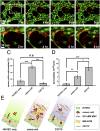
Cancer cell extravasation. (A) Extravasation of cancer cells (red) introduced into the vascular network (HUVECs, green) is monitored in real time. (B) Magnified images of white dotted box in A show extravasation of cancer cells. (C) Percent of cancer cells extravasated varies significantly among the vascular networks embedded in different microenvironments, i.e., acellular and bone or muscle-mimicking microenvironment, respectively. (D) Permeability values increased when cells are added to mimic the two organ-specific microenvironments compared with HUVEC only condition. (E) Schematic of HUVEC only, osteo-cell, and C2C12 cell added systems. HUVECs are shown as green cells that form vessel, osteo-cells are blue colored cells and secrete bone matrix as shown in yellow, and C2C12 cells are depicted as yellow cells. Cancer cells are colored in red and seen both in the vessels as well as extravasated out in the surrounding matrix.
Fig. 3.
Fig. 3.
Percent of cancer cell extravasation and vascular permeability in bone and muscle-mimicking microenvironment with addition of stimulating or blocking molecules. Cancer cells express the A3 adenosine receptor (A), whereas C2C12 embedded matrices secrete adenosine as shown by MS data (268 m/z peak) (B) and CD73 immunofluorescent staining (C). Percentage of cancer cells that extravasate (D) and permeability of the vasculature (F) in the BMi microenvironment with OD hBM-MSCSs, with and without adenosine. Extravasation rate decreased significantly with addition of adenosine, whereas the permeability increased with adenosine. Percentage of cancer cells that extravasate (E) and permeability of the vasculature (G) in the muscle-mimicking microenvironment with C2C12 cells, with and without PSB10. Although blocking of A3AR with the addition of PSB10 did not alter the permeability of the vasculature, cancer cell extravasation rate increased significantly. (H) Schematic of osteo-cell and C2C12 cell added systems with adenosine, A3 adenosine receptor and its antagonist PSB10.
Fig. 4.
Fig. 4.
Cancer cell extravasation and endothelial cell (EC) permeability change in the presence of flow through the vasculature. Extravasation of cancer cells (A) and permeability of the vasculature (B) decreased significantly with the addition of flow. (C) Extravasated cancer cells migrated further in the flow condition vs. the static condition. Actin (yellow) within ECs in static condition (D) and under conditions when flow was added in the vasculature (E). DAPI (nucleus, blue).

References

    1. Valastyan S, Weinberg RA. Tumor metastasis: Molecular insights and evolving paradigms. Cell. 2011;147(2):275–292. - PMC - PubMed
    1. Chambers AF, Groom AC, MacDonald IC. Dissemination and growth of cancer cells in metastatic sites. Nat Rev Cancer. 2002;2(8):563–572. - PubMed
    1. Hanahan D, Weinberg RA. Hallmarks of cancer: The next generation. Cell. 2011;144(5):646–674. - PubMed
    1. Fidler IJ. The pathogenesis of cancer metastasis: The ‘seed and soil’ hypothesis revisited. Nat Rev Cancer. 2003;3(6):453–458. - PubMed
    1. Bussard KM, Gay CV, Mastro AM. The bone microenvironment in metastasis: What is special about bone? Cancer Metastasis Rev. 2008;27(1):41–55. - PubMed

Publication types