Comparative Study
. 2015 Feb;35(2):421-9.
doi: 10.1161/ATVBAHA.114.304881. Epub 2014 Dec 18.

Molecular controls of lymphatic VEGFR3 signaling

Affiliations
Comparative Study

Molecular controls of lymphatic VEGFR3 signaling

Yong Deng et al. Arterioscler Thromb Vasc Biol. 2015 Feb.

Abstract

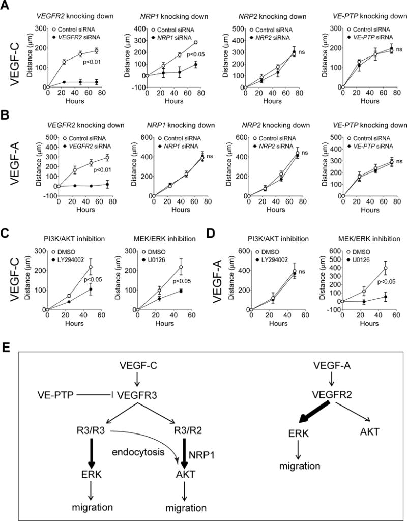
Objectives: Vascular endothelial growth factor receptor 3 (VEGFR3) plays important roles both in lymphangiogenesis and angiogenesis. On stimulation by its ligand VEGF-C, VEGFR3 is able to form both homodimers as well as heterodimers with VEGFR2 and activates several downstream signal pathways, including extracellular signal-regulated kinases (ERK)1/2 and protein kinase B (AKT). Despite certain similarities with VEGFR2, molecular features of VEGFR3 signaling are still largely unknown.

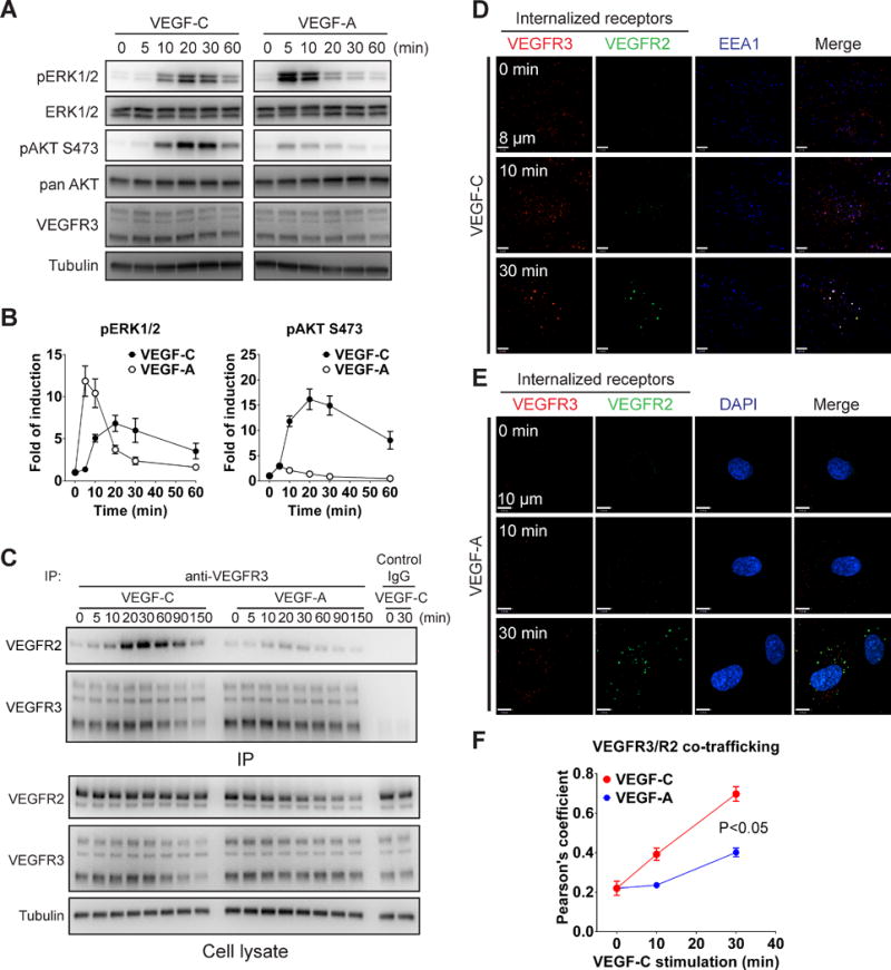
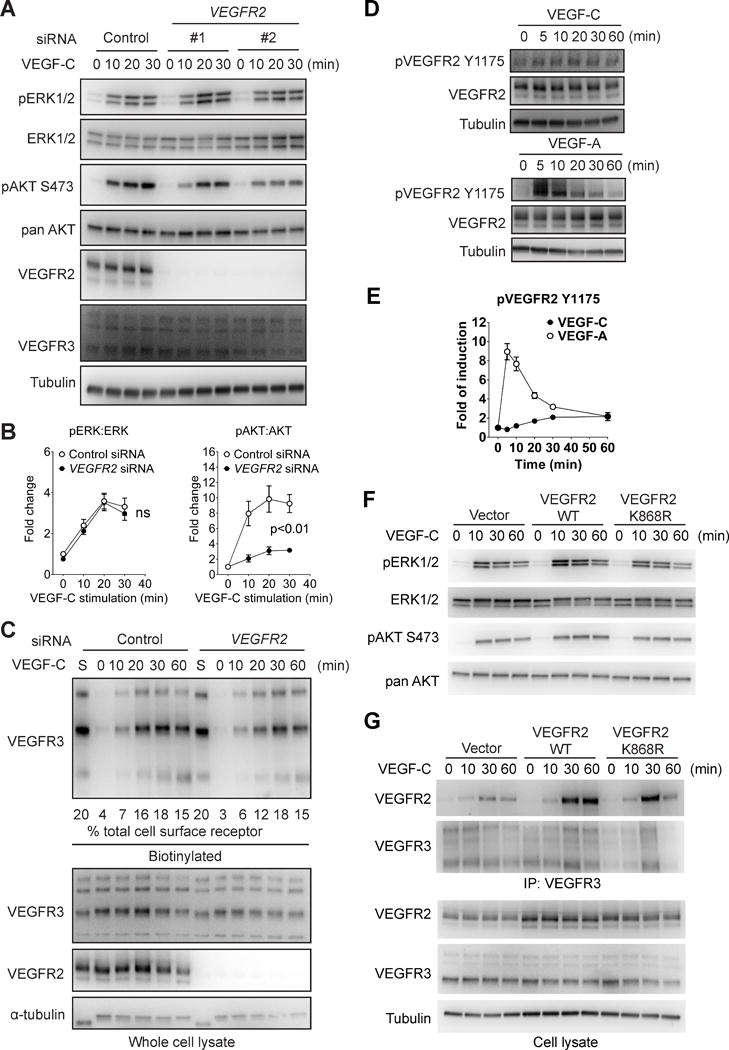
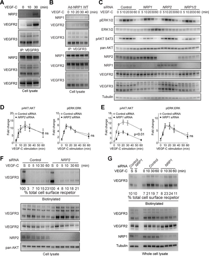
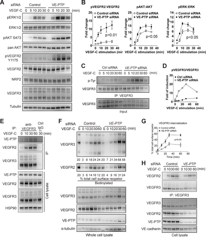
Approach and results: Human dermal lymphatic endothelial cells were used to examine VEGF-C-driven activation of signaling. Compared with VEGF-A activation of VEGFR2, VEGF-C-induced VEGFR3 activation led to a more extensive AKT activation, whereas activation of ERK1/2 displayed a distinctly different kinetics. Furthermore, VEGF-C, but not VEGF-A, induced formation of VEGFR3/VEGFR2 complexes. Silencing VEGFR2 or its partner neuropilin 1 specifically abolished VEGF-C-induced AKT but not ERK activation, whereas silencing of neuropilin 2 had little effect on either signaling pathway. Finally, suppression of vascular endothelial phosphotyrosine phosphatase but not other phosphotyrosine phosphatases enhanced VEGF-C-induced activation of both ERK and AKT pathways. Functionally, both ERK and AKT pathways are important for lymphatic endothelial cells migration.

Conclusions: VEGF-C activates AKT signaling via formation of VEGFR3/VEGFR2 complex, whereas ERK is activated by VEGFR3 homodimer. Neuropilin 1 and vascular endothelial phosphotyrosine phosphatase are involved in regulation of VEGFR3 signaling.

Keywords: NRP1; NRP2; VE-PTP; VEGF-C; VEGFR2; VEGFR3.


Conflict of interest statement

Disclosure: The authors have declared that no conflict of interest exists.

Figures

Figure 1
Figure 1. Comparison of VEGFR2 and VEGFR3 signaling
(A) Serum-starved HDLECs were stimulated with VEGF-C (100 ng/ml) or VEGF-A (50 ng/ml) and activation of ERK1/2 and AKT was determined by Western blotting. (B) Quantitative analyses of ERK and AKT phosphorylation in panel A normalized to total ERK and AKT levels, respectively. Data represent mean±SEM of three independent experiments. (C) Serum-starved HDLECs were stimulated with 100 ng/ml VEGF-C or 50 ng/ml VEGF-A for the indicated length of time. VEGFR3 immunoprecipitate was then probed with anti-VEGFR2 antibody (upper panel). Lower panel: Western blot analysis of total cell lysates corresponding to the upper panel. (D–E) Serum-starved HDLECs were incubated with anti-VEGFR3 and VEGFR2 antibodies and then stimulated with 100 ng/ml VEGF-C (D) or 50 ng/ml VEGF-A (E) for the indicated length of time. Internalized VEGFR3 (red) and VEGFR2 (green) were visualized by confocal microscopy. Early endosome was labeled with anti- early endosome antigen 1 (EEA1) (D, blue) and nuclear with DAPI (E, blue). (F) Co-localization of VEGFR3 with VEGFR2 was quantified using Pearson’s statistics. At least six fields of more than twenty cells were used for quantification. Statistical analysis was performed using two way’s ANOVA.
Figure 2
Figure 2. VEGFR2 is involved in VEGF-C-induced AKT but not ERK activation
(A) HDLECs were transfected with two different siRNAs targeting human VEGFR2 or a negative control siRNA. The cells were then serum-starved and stimulated with 100 ng/ml VEGF-C. Activation of ERK1/2 and AKT was examined using Western blotting. (B) Quantitative analyses of ERK and AKT phosphorylation. Data represents Mean±SEM of three independent experiments with three distinct VEGFR2 siRNA sequences. Statistical analysis was performed using two way’s ANOVA. (C) HDLECs were biotin labeled as described in the Methods section and VEGF-C-induced VEGFR3 internalization in HDLECs treated with VEGFR2 or control siRNAs was determined by Western blotting of cell lysates at indicated time points (upper panel). Quantification was accomplished by deriving the percentage of internalized VEGFR3 to the amount of VEGFR3 present on the cell surface prior to VEGF-C treatment (lane “S”). Note that only 20% of lane S sample is loaded in the blot shown. Numbers under the blot refer to the % fraction of VEGFR3 internalized at various time points after VEGF-C stimulation. Note the absence of significant effect of VEGFR2 knockdown on VEGFR3 internalization. (D) Serum-starved HDLECs were stimulated with 100 ng/mL VEGF-C and VEGFR2 Y1175 phosphorylation was determined as shown. (E) Quantitative analyses of VEGFR2 Y1175 phosphorylation normalized to total VEGFR2 protein level. Data represents Mean±SEM of three independent experiments. (F) HDLECs infected with lentiviruses expressing either wild type VEGFR2, kinase-dead VEGFR2 mutant (K868R) or an empty control virus were stimulated with 100 ng/ml VEGF-C and activation of ERK and AKT was examined by Western blotting as indicated. (G) Upper panel: Western blot analysis of VEGFR3 antibody immunoprecipitate from HDLECs transduced with wild type or kinase-dead (K868R) VEGFR2 constructs or an empty virus control. Lower panel: Western blot analysis of total cell lysates corresponding to the upper panel.
Figure 3
Figure 3. NRP1 but not NRP2 is required for VEGF-C signaling
(A, B) Serum-starved HDLECs were stimulated with 100 ng/ml VEGF-C and then subjected to Western blotting with anti-NRP2 (A) or NRP1 (B) antibodies of VEGFR3 antibody immunoprecipitates (upper panels). Lower panels: Western blotting of whole cell lysates. (C) HDLECs transfected with NRP1, NRP2 or control siRNAs were serum-starved and stimulated with 100 ng/ml VEGF-C. Activation of VEGFR3 signaling was examined by Western blotting as indicated. (D–E) Quantification of ERK and AKT phosphorylation after NRP2 (D) or NRP1 (E) knockdowns. Data represents Mean±SEM of three independent experiments. Statistical analysis was performed using two way’s ANOVA. (F–G) VEGF-C-induced VEGFR3 internalization following NRP2 (F) or NRP1 (G) knocked down and control HDLECs was determined by serial Western blotting of cell lysates after cell surface biotinylation as described in Methods. siRNAs was determined by Western blotting of cell lysates at indicated time points (upper panel). Quantification was accomplished by deriving the percentage of internalized VEGFR3 to the amount of VEGFR3 present on the cell surface prior to VEGF-C treatment (lane “S”). Note that 100% of lane S sample was loaded in Panel F and 10% of lane S loaded in panel G. The numbers below the blots refer to percentage of internalized VEGFR3.
Figure 4
Figure 4. VE-PTP negatively regulates VEGFR3 signaling and endocytosis
(A) HDLECs transfected with VE-PTP or control siRNAs were serum-starved and stimulated with 100 ng/ml VEGF-C. Activation of VEGFR3 signaling was examined by Western blotting as indicated. (B) Quantitative analyses of ERK, AKT and VEGFR2 phosphorylation shown in panel A. Data represents mean±SEM of three independent experiments. Statistical analysis was performed using two way’s ANOVA. (C) HDLECs transfected with VE-PTP or control siRNAs were serum-starved and stimulated with 100 ng/ml VEGF-C. VEGFR3 tyrosine phosphorylation was examined by blotting of VEGFR3 antibody immunoprecipitate with anti-tyrosine antibody. (D) Quantification of panel C. Data represents mean±SEM of three independent experiments. (E) Serum-starved HDLECs were stimulated with 100 ng/ml VEGF-C and then VEGFR3/VE-PTP interaction was examined by Western blotting with anti-VE-PTP antibody of VEGFR3 immunoprecipitate. Ctrl IgG: negative control. (F–G) Surface biotinylation assay analysis of VEGF-C-induced VEGFR3 internalization following HDLEC treatment with VE-PTP or control siRNAs. Upper panel: Western blot analysis of internalized VEGFR3. Lower panel: Western blots of total cell lysates corresponding to the upper panel. Quantification was performed as described in Fig 3F,G. Note that 20% of lane S sample was loaded. Data in G is a summary of the quantification and represented mean±SEM of at least three independent experiments. Statistical analysis was performed using two way’s ANOVA. (H) Serum-starved HDLECs treated with anti-VE-PTP or control siRNAs were stimulated with 100 ng/ml VEGF-C followed by Western blotting with anti-VEGFR2 antibody of VEGFR3 immunoprecipiates. VEGFR3/VEGFR2 interaction was determined by IP using antibodies against VEGFR3 and western blot.
Figure 5
Figure 5. VEGFR2 and NRP1 but not VE-PTP or NRP2 are required for VEGF-C-induced cell migration
(A–B) Effect of knockdown of VEGFR2, NRP1, NRP2 and VE-PTP on VEGF-C- (A) and VEGF-A- (B) induced migration of HDLECs. (C–D) Effect of inhibition of MEK/ERK or PI3K/AKT pathways on VEGF-C- (C) and VEGF-A- (D) induced migration of HDLECs. In A-D, data represent the results of the distance with VEGF stimulation minus that without VEGF. Therefore they specifically show VEGF-induced migration. Data in A–D represent mean±SEM of at least four independent experiments. Statistical analyses were performed using two way’s ANOVA. (E) A schematic model of VEGF-C/VEGFR3 signaling.

Similar articles

Cited by

References

    1. Shibuya M. Vascular endothelial growth factor and its receptor system: Physiological functions in angiogenesis and pathological roles in various diseases. Journal of biochemistry. 2013;153:13–19. - PMC - PubMed
    1. Koch S, Tugues S, Li X, Gualandi L, Claesson-Welsh L. Signal transduction by vascular endothelial growth factor receptors. Biochem J. 2011;437:169–183. - PubMed
    1. Alitalo K. The lymphatic vasculature in disease. Nat Med. 2011;17:1371–1380. - PubMed
    1. Potente M, Gerhardt H, Carmeliet P. Basic and therapeutic aspects of angiogenesis. Cell. 2011;146:873–887. - PubMed
    1. Benedito R, Rocha SF, Woeste M, Zamykal M, Radtke F, Casanovas O, et al. Notch-dependent vegfr3 upregulation allows angiogenesis without vegf-vegfr2 signalling. Nature. 2012;484:110–114. - PubMed

Publication types

MeSH terms

Substances