

Peptide internalization enabled by folding: triple helical cell-penetrating peptides
- PMID: 25524829
- PMCID: PMC4430089
- DOI: 10.1002/psc.2725
Peptide internalization enabled by folding: triple helical cell-penetrating peptides
Abstract
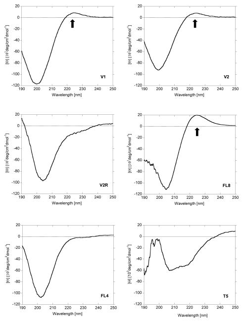
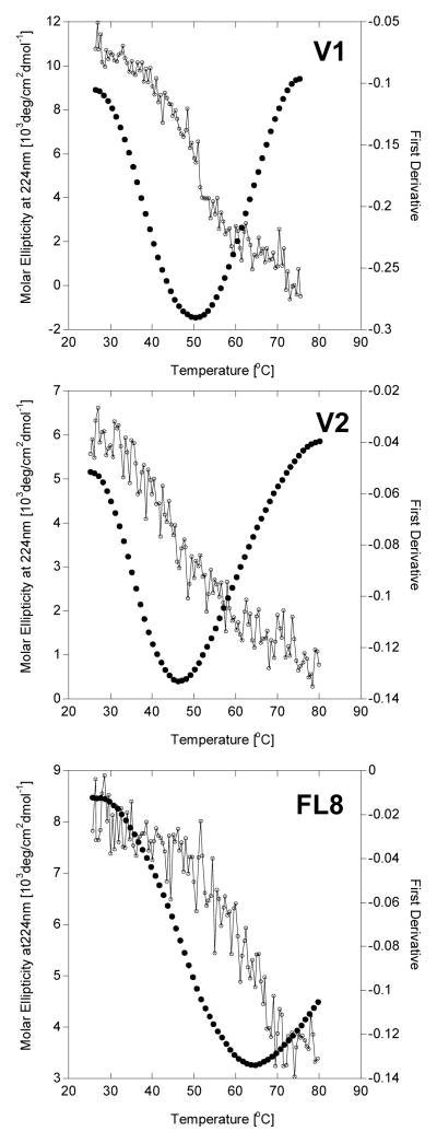
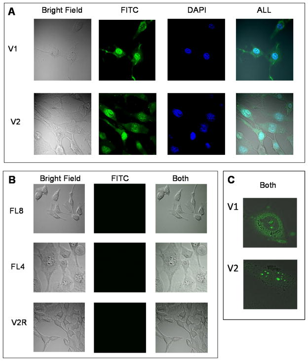
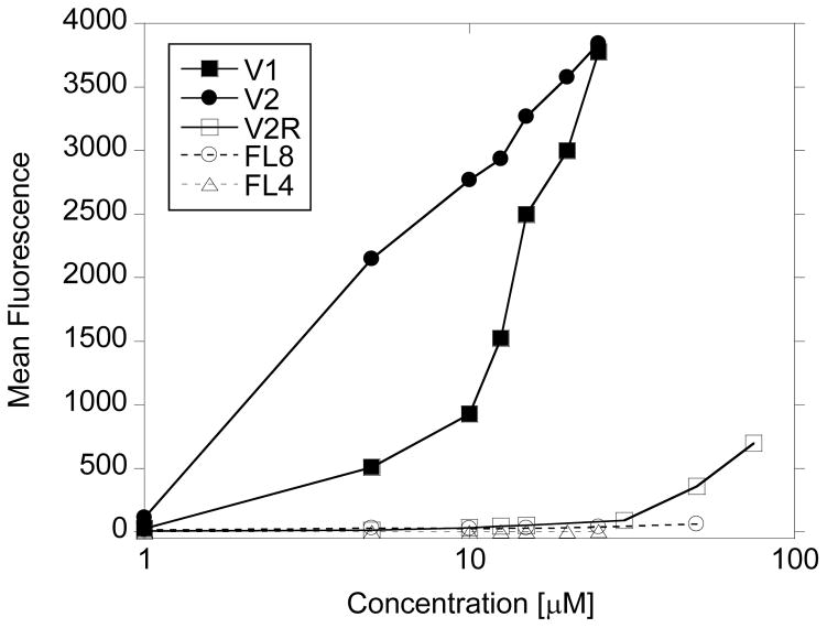
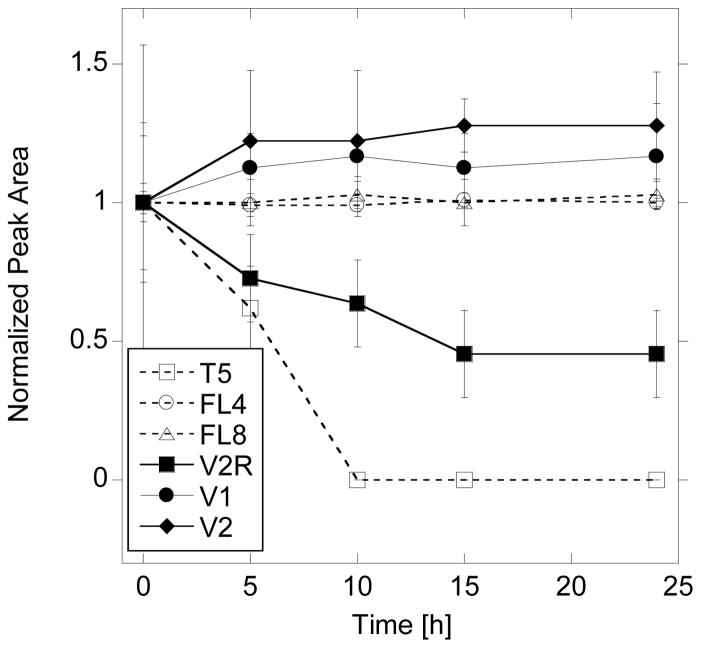
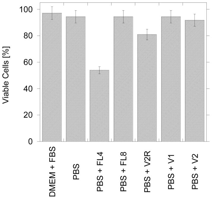
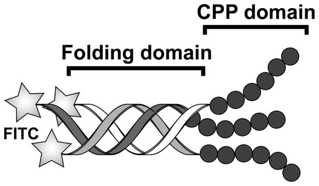
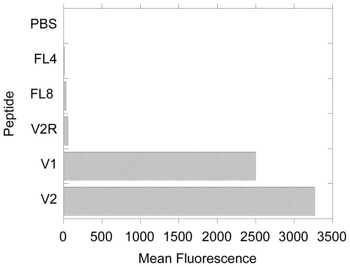
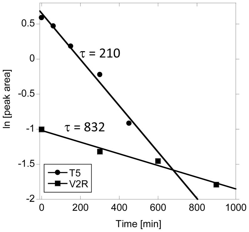
Cell-penetrating peptides (CPPs) are known as efficient transporters of molecular cargo across cellular membranes. Their properties make them ideal candidates for in vivo applications. However, challenges in the development of effective CPPs still exist: CPPs are often fast degraded by proteases and large concentration of CPPs required for cargo transporting can cause cytotoxicity. It was previously shown that restricting peptide flexibility can improve peptide stability against enzymatic degradation and limiting length of CPP peptide can lower cytotoxic effects. Here, we present peptides (30-mers) that efficiently penetrate cellular membranes by combining very short CPP sequences and collagen-like folding domains. The CPP domains are hexa-arginine (R6) or arginine/glycine (RRGRRG). Folding is achieved through multiple proline-hydroxyproline-glycine (POG [proline-hydroxyproline-glycine])n repeats that form a collagen-like triple helical conformation. The folded peptides with CPP domains are efficiently internalized, show stability against enzymatic degradation in human serum and have minimal toxicity. Peptides lacking correct folding (random coil) or CPP domains are unable to cross cellular membranes. These features make triple helical cell-penetrating peptides promising candidates for efficient transporters of molecular cargo across cellular membranes.
Keywords: cell-penetrating peptides; collagen peptides; enzymatic degradation; internalization; intracellular delivery; triple helix.
Copyright © 2014 European Peptide Society and John Wiley & Sons, Ltd.
Figures
References
-
- Wang B, Siahaan TJ, Soltero AR, editors. Drug Delivery: Principles and Applications. John Wiley & Sons Inc; Hoboken, NJ: 2005.
-
- Gu Z, Biswas A, Zhaoab M, Tang Y. Tailoring nanocarriers for intracellular protein delivery. Chem Soc Rev. 2011;40:3638–3655. - PubMed
-
- Khan DR. The use of nanocarriers for drug delivery in cancer therapy. J Cancer Sci & Therapy. 2010;2:58–62.
-
- Lindgren M, Hällbrink M, Prochiantz A, Langel U. Cell-penetrating peptides. Trends Pharmacol Sci. 2000;21:99–103. - PubMed
-
- Wang F, Wanga Y, Zhanga X, Zhanga W, Guo S, Jin F. Recent progress of cell-penetrating peptides as new carriers for intracellular cargo delivery. J Controlled Release. 2014;174:126–136. - PubMed
Publication types
MeSH terms
Substances
Grants and funding
LinkOut - more resources
Full Text Sources
Other Literature Sources