. 2015 Jul 1;212(1):95-105.
doi: 10.1093/infdis/jiu813. Epub 2014 Dec 18.

Host-pathogen Interaction at the Intestinal Mucosa Correlates With Zoonotic Potential of Streptococcus suis

Affiliations

Host-pathogen Interaction at the Intestinal Mucosa Correlates With Zoonotic Potential of Streptococcus suis

Maria Laura Ferrando et al. J Infect Dis. .

Abstract

Background: Streptococcus suis has emerged as an important cause of bacterial meningitis in adults. The ingestion of undercooked pork is a risk factor for human S. suis serotype 2 (SS2) infection. Here we provide experimental evidence indicating that the gastrointestinal tract is an entry site of SS2 infection.

Methods: We developed a noninvasive in vivo model to study oral SS2 infection in piglets. We compared in vitro interaction of S. suis with human and porcine intestinal epithelial cells (IEC).

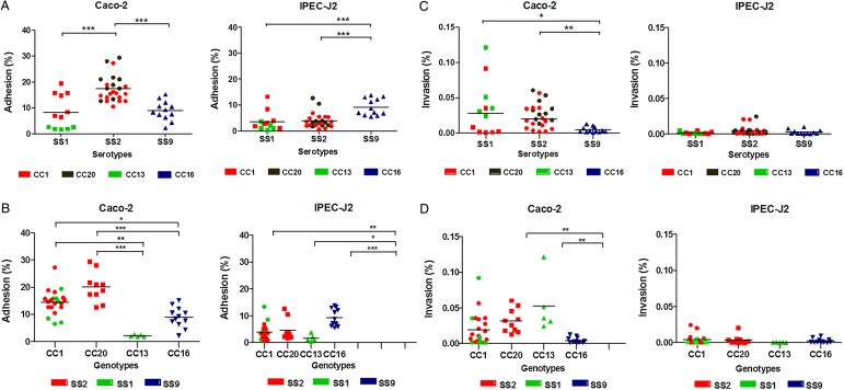
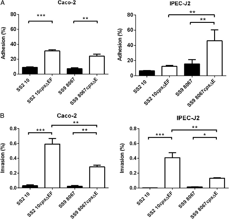
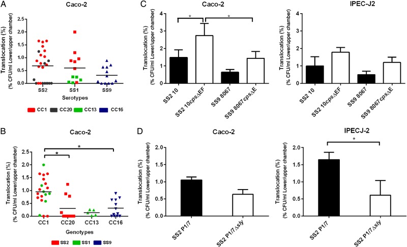
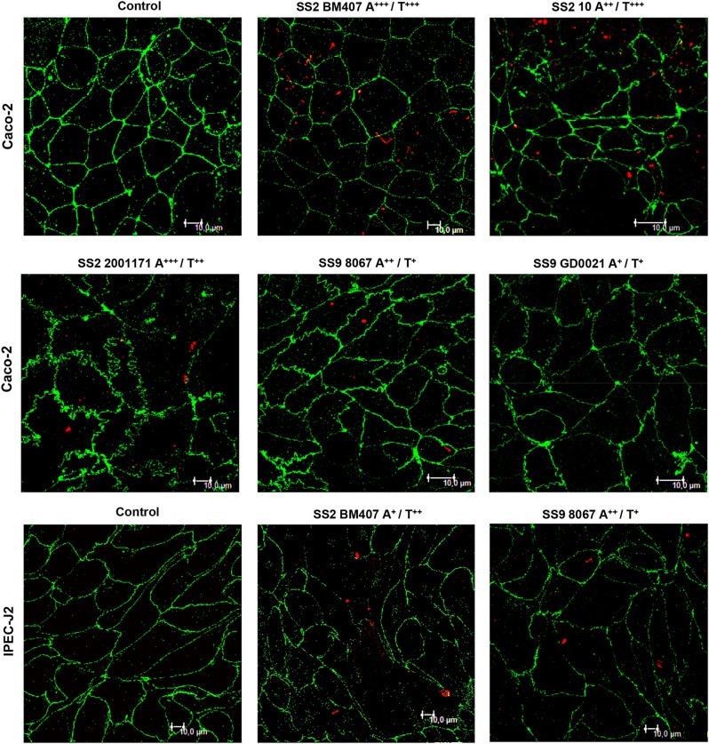
Results: Two out of 15 piglets showed clinical symptoms compatible with S. suis infection 24-48 hours after ingestion of SS2. SS2 was detected in mesenteric lymph nodes of 40% of challenged piglets. SS2 strains isolated from patients showed significantly higher adhesion to human IEC compared to invasive strains isolated from pigs. In contrast, invasive SS9 strains showed significantly higher adhesion to porcine IEC. Translocation across human IEC, which occurred predominately via a paracellular route, was significantly associated with clonal complex 1, the predominant zoonotic genotype. Adhesion and translocation were dependent on capsular polysaccharide production.

Conclusions: SS2 should be considered a food-borne pathogen. S. suis interaction with human and pig IEC correlates with S. suis serotype and genotype, which can explain the zoonotic potential of SS2.

Keywords: Streptococcus suis; clonal complex; intestinal translocation; piglets; serotype; tight junctions; zoonotic infections.


Figures

Figure 1.
Figure 1.
S. suis serotype 2 (SS2) in intestinal mucosa tissue derived from SS2 free piglets after oral SS2 challenge. Multi-spectral imaging of immunohistochemistry staining of intestinal tissue of piglet 1 (not infected, jejunum) and piglet 11 (jejunum and colon). Sections were stained with hematoxylin 1×. SS2 was detected using rabbit monoclonal anti-SS2 antibody. Slides were evaluated by light microscopy using 2.5×, 20× and 63× immersion oil objectives in digital bright field image and digital fluorescence (Nuance software).
Figure 2.
Figure 2.
S. suis strains associated with different ability to adhere to and invade the human and porcine IEC. A and B, Multiple comparisons of S. suis strains with different serotypes (SS) (A) and genotypes (CC) (B) for their adhesion to human (Caco-2) and porcine (IPEC-J2) EIC. C and D, Multiple comparisons of S. suis strains with different serotypes (SS) (C) and genotypes (CC) (D) for their invasion capacity to Caco-2 and IPEC-J2 cells. Serotypes analyzed: SS1 ▪, SS2 • and SS9 ▴. MLST Clonal Complex (CC) CC1, CC20, CC13 and CC16 strains are indicated in red, grey, green, and blue respectively. Genotypes analyzed: CC1 •, CC20 ▪, CC13 ▴, and CC16 ▾. SS2, SS9 and SS1 strains are indicated in red, blue and green, respectively. Lines denote the median percentage of adhesion for each group of strains. *P < .05; **P < .01; ***P < .001 (Kruskal–Wallis test).
Figure 3.
Figure 3.
The polysaccharide capsule hampers S. suis adhesion to and invasion of human and porcine IEC. A, Adhesion and (B) invasion of Caco-2 and IPEC-J2 cells displayed by the wild type (WT) strains SS2 strain 10 and SS9 strain 8067 and their isogenic unencapsulated mutants 10cpsΔEF and 8067ΔcpsΔE respectively. Data are given as means ± standard error of the mean (SEM) of at least 2 separate experiments performed in triplicate and analyzed by 1-way analysis of variance (ANOVA). *P < .05; **P < .001.
Figure 4.
Figure 4.
Genetic background of S. suis determines the translocation across human IEC. A, Multiple comparisons of S. suis translocation across differentiated Caco-2 monolayers after 6 h of co-incubation, analyzed by S. suis serotype (SS): SS1 ▪, SS2 • and SS9 ▴. MLST Clonal Complex (CC) CC1, CC20, CC13 and CC16 strains are indicated in red, grey, green, and blue respectively. B, Multiple comparisons of S. suis translocation across differentiated Caco-2 monolayers after 6 h of co-incubation, analyzed by S. suis genotype: MLST clonal complex (CC) CC1 •, CC20 ▪, CC13 ▴, and CC16 ▾. SS2, SS9 and SS1 strains are indicated in red, blue, and green, respectively. Lines denote the median percentage of translocation for each group of strains, analyzed by Kruskal–Wallis test. C, Comparison of bacterial translocation efficiency of unencapsulated (10cpsΔEF and 8067ΔcpsΔE) mutant strains and their wild-type parent strains across differentiated human (Caco-2) and porcine (IPEC-J2) IEC. D, Comparison of bacterial translocation efficiency of a suilysin mutant (P1/7Δsly) and its wild-type parent strain across differentiated human (Caco-2) and porcine (IPEC-J2) IEC. Data are given as means ± standard error of the mean (SEM) of at least 2 independent experiments, each performed in triplicate and analyzed by 1-way analysis of variance (ANOVA). *P < .05; **P < .01; ***P < .001.
Figure 5.
Figure 5.
S. suis strains with high translocation capacity induce re-arrangement of Tight Junction (TJ) of Caco-2 and IPEC-J2 cells. Confocal imaging of A, Caco-2 and B, IPEC-J2 intestinal epithelial cells stained for TJ protein occludin (green) in the absence (control) and in the presence of S. suis strains with low (+) medium (++), high (+++),adhesion (A) and translocation (T) ability, after 4 h of co-incubation (see “Materials and Methods” section for definitions of low, medium, and high). Bacteria (red) were stained with rabbit monoclonal antisera raised against capsular polysaccharides of serotype 2 and were counterstained with Alexa Fluor 488–conjugated anti-rabbit antibodies. Co-localization of bacteria and TJ (yellow). Bar 10 µm.
Figure 6.
Figure 6.
S. suis co-localizes with cellular junctions and translocates from the apical to the basolateral side of polarized IEC. A, 3D confocal images of Caco-2 and IPEC-J2 cell layers stained for the TJ protein occludin (green) in the absence (control) and in the presence of S. suis 2 BM407 (red) after 4 h incubation. The nuclei of intestinal cells are stained with DAPI (blue). Lower and side strips are relative to x and y sections through the monolayer where Z-stack images are made from the apical (0 µm) to the basolateral (180 µm) side. Bacteria stained in light pink indicate co-localization with occludin (black arrow). B, TEM of differentiated Caco-2 cells fixed at 4 h after infection with S. suis serotype 2 strain BM407. SS2 strain BM407 adheres to the apical microvilli after apical infection (yellow arrows). Translocating bacteria (red arrows) are found in close vicinity to tight junctions and within a vacuole (*). Images were made at magnifications of 2500×, 5000× and 10 000×. Bar: 10, 5 or 2 µm as indicated.

References

    1. Wertheim HF, Nghia HD, Taylor W, Schultsz C. Streptococcus suis: an emerging human pathogen. Clin Infect Dis 2009; 48:617–25. - PubMed
    1. Huong VT, Ha N, Huy NT, et al. Epidemiology, clinical manifestations, and outcomes of Streptococcus suis infection in humans. Emerg Infect Dis 2014; 20:1105–14. - PMC - PubMed
    1. Nghia HD, Tu le TP, Wolbers M, et al. Risk factors of Streptococcus suis infection in Vietnam. A case-control study. PLoS One 2011; 6:e17604. - PMC - PubMed
    1. Takeuchi D, Kerdsin A, Pienpringam A, et al. Population-based study of Streptococcus suis infection in humans in Phayao Province in northern Thailand. PLoS One 2012; 7:e31265. - PMC - PubMed
    1. Williams DM, Lawson GH, Rowland AC. Streptococcal infection in piglets: the palatine tonsils as portals of entry for Streptococcus suis. Res Vet Sci 1973; 15:352–62. - PubMed

Publication types