. 2014 Dec 18;56(1):177-84.
doi: 10.1167/iovs.14-15550.

Pioglitazone restores IGFBP-3 levels through DNA PK in retinal endothelial cells cultured in hyperglycemic conditions

Affiliations

Pioglitazone restores IGFBP-3 levels through DNA PK in retinal endothelial cells cultured in hyperglycemic conditions

Shalini Thakran et al. Invest Ophthalmol Vis Sci. .

Abstract

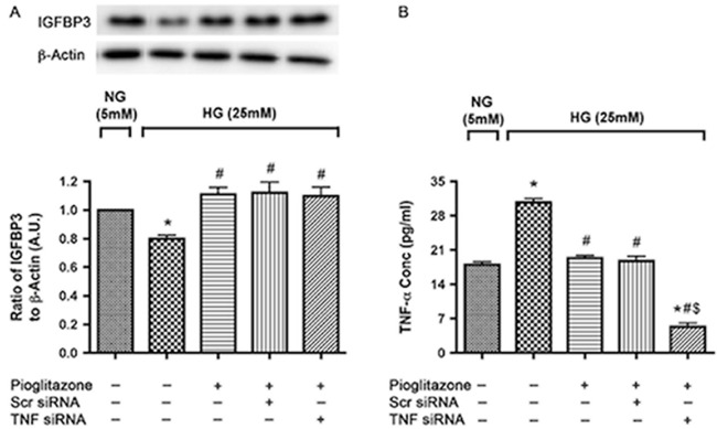
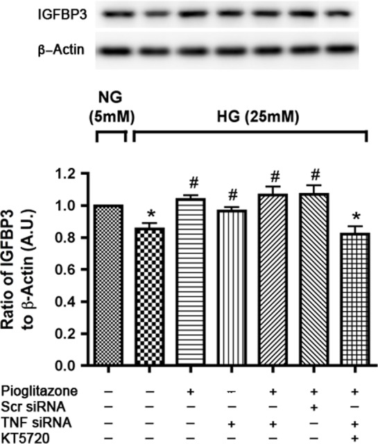
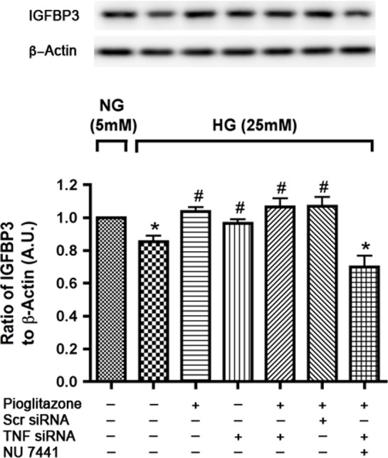
Purpose: Previously, we reported that pioglitazone prevented insulin resistance and cell death in type 2 diabetic retina by reducing TNFα and suppressor of cytokine signaling 3 (SOCS3) levels. Numerous reports suggest prominent vasoprotective effects of insulin growth factor binding protein-3 (IGFBP-3) in diabetic retinopathy. We hypothesized that pioglitazone protects against retinal cell apoptosis by regulating IGFBP-3 levels, in addition to reducing TNFα. The current study explored potential IGFBP-3 regulatory pathways by pioglitazone in retinal endothelial cells cultured in high glucose.

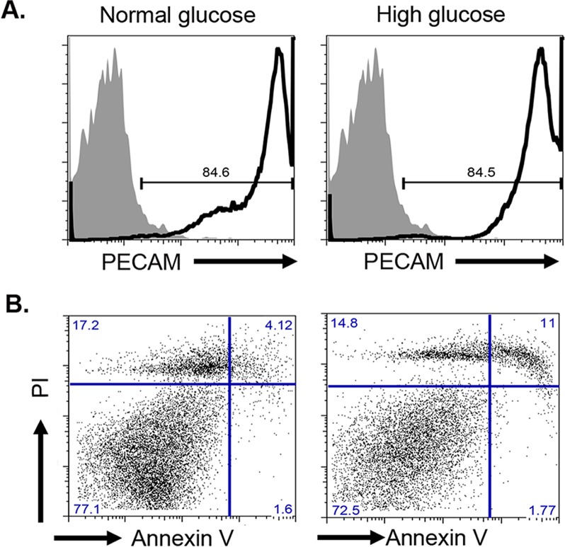
Methods: Primary human retinal endothelial cells (REC) were grown in normal (5 mM) and high glucose (25 mM) and treated with pioglitazone for 24 hours. Cell lysates were processed for Western blotting and ELISA analysis to evaluate IGFBP-3, TNFα, and cleaved caspase 3 protein levels.

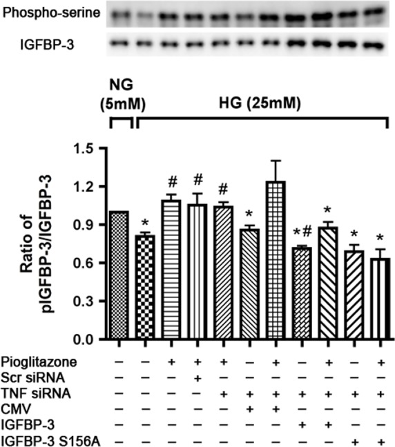
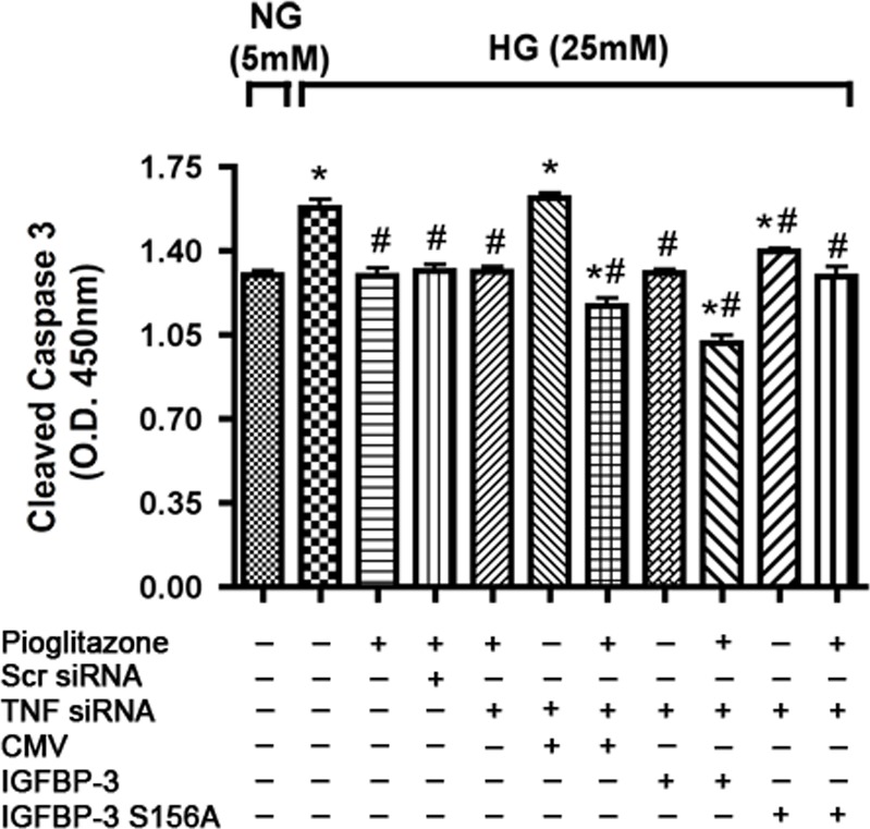
Results: Our results show that treatment with pioglitazone restored the high glucose-induced decrease in IGFBP-3 levels. This regulation was independent of TNFα actions, as reducing TNFα levels with siRNA did not prevent pioglitazone from increasing IGFBP-3 levels. Pioglitazone required protein kinase A (PKA) and DNA-dependent protein kinase (DNA PK) activity to regulate IGFBP-3, as specific inhibitors for each protein prevented pioglitazone-mediated normalization of IGFBP-3 in high glucose. Insulin growth factor binding protein-3 activity was increased and apoptosis decreased by pioglitazone, which was eliminated when serine site 156 of IGFBP-3 was mutated suggesting a key role of this phosphorylation site in pioglitazone actions.

Conclusions: Our findings suggest that pioglitazone mediates regulation of IGFBP-3 via activation of PKA/DNA PK pathway in hyperglycemic retinal endothelial cells.

Keywords: DNA PK; IGFBP-3; pioglitazone; retinal endothelial cells.


Figures

Figure 1
Figure 1
High glucose-induced REC cell death. (A) Flow cytometry analysis of PECAM-1 in REC. Solid histogram shows levels of mouse IgG1κ isotype control and open histogram shows experimental sample results. (B) Annexin V versus PI labeling to determine apoptosis. Normal and high glucose–cultured cells were labeled with Annexin V-FITC and PI prior analysis. Percentage dead cells: percent Annexin V+PI+, percent live cells Annexin VnegPIneg.
Figure 2
Figure 2
Pioglitazone induced IGFBP-3 levels in high-glucose medium in a TNFα independent way. (A) Western blot analysis of IGFBP-3 to β-actin ratio in REC transfected with scrambled and TNFα siRNA for 24 hours followed by treatment with 25 μM pioglitazone for 24 hours in 5 and 25 mM glucose. (B) Bar graph of TNFα levels after TNFα transfection. (A) *P < 0.05 versus untreated NG control. #P < 0.05 versus untreated HG control. N = 4. (B) *P < 0.05 versus untreated NG control. #P < 0.05 versus untreated HG control. $P < 0.05 versus respective scrambled siRNA control. Data are mean ± SEM. N = 4.
Figure 3
Figure 3
Pioglitazone induced IGFBP-3 expression is through PKA activation in high-ambient glucose. Figure shows bar graph of IGFBP-3 to β-actin levels measured by Western blot in REC cultured in high glucose (25 mM) with TNFα siRNA transfection for 24 hours and then treatment with KT 5720 for 30 minutes followed by 24 hours pioglitazone treatment. Retinal endothelial cells in normal glucose (5 mM) was used as control. *P < 0.05 versus untreated normal glucose control. #P < 0.05 versus untreated high glucose control. Data are mean ± SEM, N = 4.
Figure 4
Figure 4
Pioglitazone requires DNA-PK activity to induce IGFBP-3 in high glucose medium. Western blot analysis of IGFBP3 levels in RECs transfected with TNFα siRNA and treated with NU 7441 for 30 minutes followed by pioglitazone treatment for 24 hours in high-glucose medium (25 mM). Retinal endothelial cells in normal glucose (5 mM) were used as control. *P < 0.05 versus untreated normal glucose control. #P < 0.05 versus untreated high glucose control. Data are mean ± SEM, N = 4.
Figure 5
Figure 5
Pioglitazone does not activate IGFBP-3 with transfection of IGFBP-3 S156A plasmid in high glucose medium. Bar graph of ratio of Western blots analysis of p-IGFBP-3 to IGFBP-3 in RECs cultured in high glucose transfected with TNFα siRNA followed by transfection of CMV, IGFBP-3, and IGFBP-3 S156A plasmid next day. Pioglitazone was added for 24 hours after another day. Retinal endothelial cells in normal glucose (5 mM) were used as control. Protein was immunoprecipitated with anti–IGFBP-3 antibody and immunoblotted using anti–IGFBP-3 and antiphosphoserine antibodies. *P < 0.05 versus untreated normal glucose control. #P < 0.05 versus untreated high glucose control. Data are mean ± SEM, N = 4.
Figure 6
Figure 6
IGFBP-3 S156A plasmid DNA transfection partially reverses inhibition of apoptosis with pioglitazone. Bar graph of cleaved caspase 3 expression of REC in normal (5 mM) and high glucose (25 mM). Retinal endothelial cells in high-glucose medium were transfected with TNFα siRNA for 24 hours followed by transfection with CMV, IGFBP-3, and IGFBP-3 S156A plasmid and subsequent treatment with pioglitazone next day for 24 hours. *P < 0.05 versus untreated normal glucose control. #P < 0.05 versus untreated high glucose control. Data are mean ± SEM, N = 4.

References

    1. Cheung N, Mitchell P, Wong TY. Diabetic retinopathy. Lancet. 2010; 376: 124–136. - PubMed
    1. Jiang Y, Zhang Q, Soderland C, Steinle JJ. TNFalpha and SOCS3 regulate IRS-1 to increase retinal endothelial cell apoptosis. Cell Signalling. 2012; 24: 1086–1092. - PMC - PubMed
    1. Steinle JJ. Retinal endothelial cell apoptosis. Apoptosis. 2012; 17: 1258–1260. - PMC - PubMed
    1. Joussen AM, Murata T, Tsujikawa A, Kirchhof B, Bursell SE, Adamis AP. Leukocyte-mediated endothelial cell injury and death in the diabetic retina. Am J Pathol. 2001; 158: 147–152. - PMC - PubMed
    1. Tawfik A, Sanders T, Kahook K, Akeel S, Elmarakby A, Al-Shabrawey M. Suppression of retinal peroxisome proliferator-activated receptor gamma in experimental diabetes and oxygen-induced retinopathy: role of NADPH oxidase. Invest Ophthalmol Vis Sci. 2009; 50: 878–884. - PubMed

Publication types

MeSH terms