Review
. 2015 Mar;88(1047):20140406.
doi: 10.1259/bjr.20140406. Epub 2014 Dec 19.

Peripheral limb vascular malformations: an update of appropriate imaging and treatment options of a challenging condition

Affiliations
Review

Peripheral limb vascular malformations: an update of appropriate imaging and treatment options of a challenging condition

H Madani et al. Br J Radiol. 2015 Mar.

Abstract

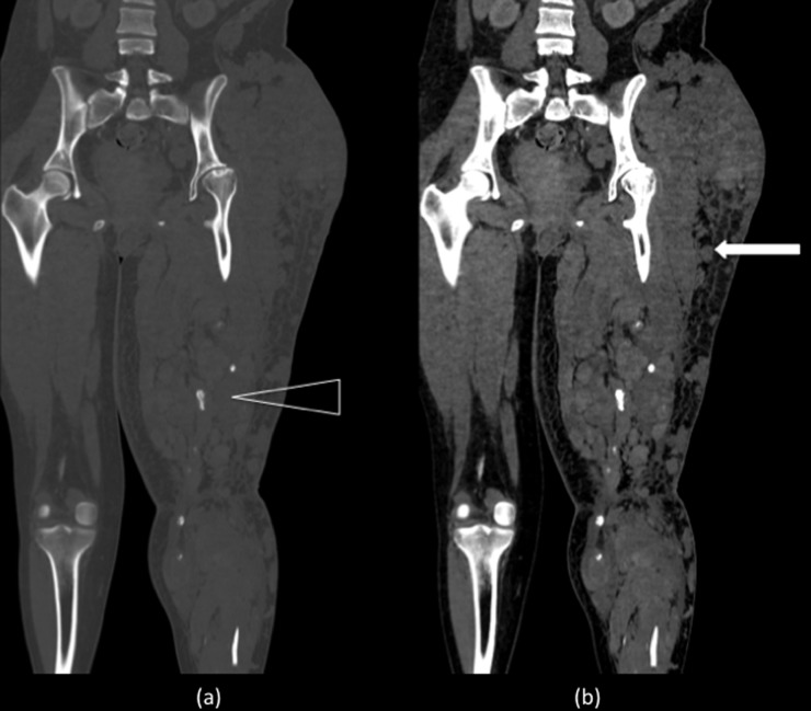
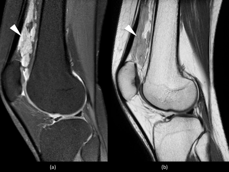
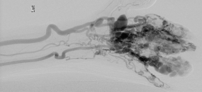
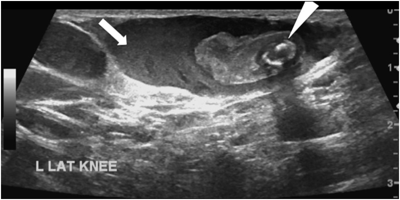
Peripheral vascular malformations encompass a wide spectrum of lesions that can present as an incidental finding or produce potentially life- or limb-threatening complications. They can have intra-articular and intraosseous extensions that will result in more diverse symptomology and present greater therapeutic challenges. Developments in classification, imaging and interventional techniques have helped to improve outcome. The onus is now placed on appropriate detailed preliminary imaging, diagnosis and classification to direct management and exclude other more common mimics. Radiologists are thus playing an increasingly important role in the multidisciplinary teams charged with the care of these patients. By fully understanding the imaging characteristics and image-guided procedures available, radiologists will be armed with the tools to meet these responsibilities. This review highlights the recent advances made in imaging and the options available in interventional therapy.


Figures

Figure 1.
Figure 1.
Plain radiographs of the left elbow demonstrate multiple calcific densities consistent with vascular phleboliths (white arrowhead) suggestive of venous malformations. There is also degenerative osteoarthritic change, with joint space loss and subtle scalloping of the radial neck (open arrowheads). These arthropathic findings are highly suggestive of a destructive arthropathy secondary to recurrent haemarthrosis.
Figure 2.
Figure 2.
Plain radiographs (a) and equivalent reconstructed three-dimensional CT image (b) of the left knee demonstrating multiple rounded calcific densities within the soft tissues, consistent with phleboliths, later confirmed to be within extensive slow-flow venous malformations.
Figure 3.
Figure 3.
Plain radiograph (a) of the left hand demonstrating lytic lesions, cortical scalloping (open arrowheads), remodelling and acro-osteolysis of the distal phalanges (white arrowhead) caused by multiple malformations, confirmed to be high-flow arteriovenous malformations on angiography (b).
Figure 4.
Figure 4.
Ultrasound of the hand, demonstrating avid colour Doppler flow features of a high-velocity arteriovenous malformation (white arrowhead), seen to be dorsal to the proximal interphalangeal joint of the little finger (open arrowhead).
Figure 5.
Figure 5.
Coronal reformat CT of the pelvis and lower limbs in bone (a) and soft tissue (b) windows showing multiple slow-flow malformations (white arrow) producing a large left thigh. The partially seen left femur is osteopenic and gracile. There are a number of phlebliths suggesting multiple venous channels (open arrowhead).
Figure 6.
Figure 6.
Sagittal short tau inversion recovery (a) and T1 weighted (b) images of the left knee showing multiple low-flow venous malformations (white arrowheads) within the supra patellar fat pad and patella femoral joint.
Figure 7.
Figure 7.
Digital subtracted angiogram of the left hand showing contrast-filled multiple dilated tortious arteriovenous malformations within in the digits, supplied by dilated radial and ulnar arteries.
Figure 8.
Figure 8.
Ultrasound image showing isoechoic and echo poor tubular structures, the largest (arrow) containing an echogenic focus representing likely thrombus with a central smaller phlebolith, with characteristic post-acoustic shadowing, pathognomonic of a venous peripheral vascular malformation (arrowhead). L Lat Knee, left lateral knee.
Figure 9.
Figure 9.
Sagittal proton density spectral pre-saturation with inversion recovery image of the knee showing a small cluster of high-signal slow-flow malformations, with the absence of flow voids, posterior to the popliteal vessels (white arrowhead). There is no intra-articular extension.
Figure 10.
Figure 10.
Coronal short tau inversion recovery image of the pelvis and upper thighs shows high-intensity heterogeneous masses involving the subcutaneous tissue and muscle surrounding a gracile left hemi pelvis and femur (black arrow). The lack of signal void indicates slow-flow venous malformations. There are numerous phleboliths lying within the vascular channels seen as signal voids in the lesions (arrowheads).
Figure 11.
Figure 11.
Coronal T1 weighted images of the knees showing multiple flow voids surrounding the right knee (white arrowhead), consistent with complex high-flow arterial malformations.
Figure 12.
Figure 12.
Coronal T1 weighted (a) and short tau inversion recovery (b) images show a large cluster of small high-flow vessels seen as signal voids (open arrowhead) within the left popliteal fossa (a), communicating superiorly with a large calibre slow-flow superficial femoral vein (white arrowheads).
Figure 13.
Figure 13.
Sagittal proton density (PD)-weighted (a) and coronal T2 weighted (b) and short tau inversion recovery (STIR) (c) sequences show multiple high-signal serpiginous structures (white arrows) within the lower thigh and intra-articular extension consistent with slow-flow peripheral venous malformations. The PD and STIR sequence demonstrate marked degenerative change within both tibiofemoral compartments. There is significant joint space narrowing with cartilage thinning, subchondral cysts (open arrowhead) (a) and bone marrow oedema (white arrowheads) (b and c).
Figure 14.
Figure 14.
Axial T1 weighted (T1W) (a), short tau inversion recovery (b) and T1W gadolinium-enhanced (c) images of the thighs showing a large well-demarcated intra-osseous extending arteriovenous malformation (AVM) (open arrowheads) (a, b) with associated surrounding enhancing anterior intramuscular lesions (white arrow), within the right thigh (c). Coronal (d) and sagittal (e) T1W gadolinium-enhanced images of the same patient, confirmed the full extent of the AVM (white arrowheads) within the diaphysis of the femur and the enhancing intramuscular lesions (white arrow) (d).
Figure 15.
Figure 15.
Digital subtraction angiogram images of a complex arteriovenous malformation surrounding the right elbow. The initial contrast run (a) confirmed the correct position of the needle (arrowhead) within the malformation, overlying the olecranon. A delayed run (b) also established a larger component of the malformation (open arrowhead). Percutaneous injection of sclerosant (c) (arrowhead), delivered as foam and air mixture, shows gradual vessel “ghosting” of the malformation.
Figure 16.
Figure 16.
Digital subtraction angiograms of high-flow arteriovenous malformations surrounding the right knee. The early contrast run (a) demonstrates filling of a hyperdynamic and enlarged superficial femoral artery (white arrowhead). Intraosseous and intra-articular components supplied by high-flow perigeniculate branches were noted (black arrowhead) (b). On the delayed phase images (c), rapid shunting was seen into the efferent dilated and partially aneurysmal veins (open arrowhead).
Figure 17.
Figure 17.
Endovascular embolization performed using liquid embolic agent (Onyx). Two medial and two lateral vessels feeding a high-flow intra-articular component were targeted with selective catherization (white arrowheads). At the end of the procedure, there was markedly reduced filling and reduced shunting with the embolic agent producing a black filling of the occluded vessels (open arrows).
Figure 18.
Figure 18.
Post-embolization confirmatory angiogram showed markedly reduced filling and shunting with expected “ghosting” artefact within the obliterated vessels in the arterial phase (white arrowhead) (a) and delayed venous phase images (black arrowhead) (b).

References

    1. Mulliken JB, Glowacki J. Hemangiomas and vascular malformations in infants and children: a classification based on endothelial characteristics. Plast Reconstr Surg 1982; 69: 412–22. - PubMed
    1. Mulliken JB, Glowacki J. Classification of pediatric vascular lesions. Plast Reconstr Surg 1982; 70: 120–1. - PubMed
    1. Legiehn GM, Heran MK. Venous malformations: classification, development, diagnosis, and interventional radiologic management. Radiol Clin North Am 2008; 46: 545–97. doi: 10.1016/j.rcl.2008.02.008 - DOI - PubMed
    1. Legiehn GM, Heran MK. Classification, diagnosis, and interventional radiologic management of vascular malformations. Orthop Clin North Am 2006; 37: 435–74. - PubMed
    1. Hyodoh H, Hori M, Akiba H, Tamakawa M, Hyodoh K, Hareyama M. Peripheral vascular malformations imaging, treatment approaches, and therapeutic issues. Radiographics 2005; 25(Suppl 1): S159–71. - PubMed

MeSH terms