. 2015 Jan;76(1):92-100.
doi: 10.1227/NEU.0000000000000552.

Cerebral hypoperfusion-assisted intra-arterial deposition of liposomes in normal and glioma-bearing rats

Affiliations

Cerebral hypoperfusion-assisted intra-arterial deposition of liposomes in normal and glioma-bearing rats

Shailendra Joshi et al. Neurosurgery. 2015 Jan.

Abstract

Background: Optimizing liposomal vehicles for targeted delivery to the brain has important implications for the treatment of brain tumors. The promise of efficient, brain-specific delivery of chemotherapeutic compounds via liposomal vehicles has yet to be achieved in clinical practice. Intra-arterial injection of specially designed liposomes may facilitate efficient delivery to the brain and to gliomas.

Objective: To test the hypothesis that cationic liposomes may be effectively delivered to both normal and glioma-bearing brain tissue utilizing a strategy of intra-arterial injection during transient cerebral hypoperfusion.

Methods: Cationic, anionic, and neutral liposomes were separately injected via the internal carotid artery of healthy rats during transient cerebral hypoperfusion. Rats bearing C6 gliomas were similarly injected with cationic liposomes. Liposomes were loaded with DilC18(5) dye whose concentrations can be measured by light absorbance and fluorescence methods.

Results: After intra-arterial injection, a robust uptake of cationic in comparison with anionic and neutral liposomes into brain parenchyma was observed by diffuse reflectance spectroscopy. Postmortem multispectral fluorescence imaging revealed that liposomal cationic charge was associated with more efficient delivery to the brain. Cationic liposomes were also readily observed within glioma tissue after intra-arterial injection. However, over time, cationic liposomes were retained longer and at higher concentrations in the surrounding, peritumoral brain than in the tumor core.

Conclusion: This study demonstrates the feasibility of cationic liposome delivery to brain and glioma tissue after intra-arterial injection. Highly cationic liposomes directly delivered to the brain via an intracarotid route may represent an effective method for delivering antiglioma agents.


Figures
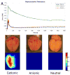
Figure. 1
Figure. 1. Representative brain liposome concentration-time curves and multispectral fluorescence images
The concentration-time curves generated by diffuse reflectance spectroscopy indicate preferential brain uptake and retention of cationic liposomes as compared to anionic and neutral liposome after intraarterial delivery (A). Post-mortem brain sample with corresponding multispectral fluorescence image shows high retention of cationic liposomes in the ipsilateral cerebral hemisphere (B). In comparison, weak signal on multispectral fluorescence images after anionic (C) and neutral liposome (D) delivery indicates poor brain targeting of these liposomes after intraarterial delivery.
Figure. 2
Figure. 2. Charge-specific regional distribution of liposomes
Diffuse reflectance spectroscopy of post mortem samples demonstrates the regional deposition of liposomes with different surface charges in specified arterial distributions. Significantly increased uptake of cationic liposomes is observed across all arterial distributions of the ipsilateral hemisphere as compared with deposition of anionic and neutral liposomes.
Figure. 3
Figure. 3. Representative concentration-time curves and post mortem multispectral fluorescence images of cationic liposomes
50% DOTAP liposomes achieved the highest peak concentration but also had the most rapid clearance. The end tissue concentration of 25% DOTAP liposomes was the same as that observed with 50% DOTAP liposomes (A). Post-mortem brain samples with corresponding multispectral fluorescence images show relatively increased retention of 50% (B) and 25% (C) DOTAP liposomes as compared to 5% (D) DOTAP liposomes which had minimal fluorescence signal.
Figure. 4
Figure. 4. Regional deposition of cationic liposomes of differing charge density
Quantitative assessment of post-mortem brain samples by diffuse reflectance spectroscopy indicates that the lowest cationic charge density liposomes (5% DOTAP) tested had the least retention across all arterial distributions. A trend toward increased retention of 25% as compared to 50% DOTAP liposomes was also seen across all arterial distributions suggesting that optimal rather than maximal cationic charge loading is necessary for efficient intrarterial delivery.
Figure. 5
Figure. 5. Distribution of cationic liposomes in glioma-bearing brain
Gross section through a C6 glioma-bearing brain is shown. Labeled (B-E) locations on the gross specimen correspond to the regions shown by confocal microscopy (A). Confocal micrographs of brain sections contralateral to tumor (B), ispilateral but remote from tumor (C), within the tumor (D), and in the peri-tumoral tissue (E) are shown. DAPI stained cell nuclei (blue) and the fluorescent liposome membrane label DiD (red) indicate liposome deposition relative to cells. Thus, high levels of liposome retention are achieved both within and in the immediate periphery of the tumor. Tu - tumor
Figure. 6
Figure. 6. Time course of cationic liposome retention in glioma-bearing brain
Gross brain section through tumor (A-C) with corresponding multispectral fluorescence images (D-F) and H&E stained sections (G-I) at 30 minutes (top row), 2 hours (middle row), and 4 hours (bottom row) after intraarterial liposome delivery are shown. High levels of fluorescence, indicative of liposome localization, are seen both within and surrounding the tumor at 30 minutes (D) and after 2 hours (E). The highest levels of fluorescence appear to be peritumoral. By 4 hours after intraarterial delivery much lower levels of fluorescence are seen within the tumor (F) although discreet areas of high intensity can be seen in regions surrounding the tumor. Tu - tumor

References

    1. Drummond DC, Noble CO, Hayes ME, Park JW, Kirpotin DB. Pharmacokinetics and in vivo drug release rates in liposomal nanocarrier development. J Pharm Sci. 2008 Mar 19; - PubMed
    1. Straubinger RM, Arnold RD, Zhou R, Mazurchuk R, Slack JE. Antivascular and antitumor activities of liposome-associated drugs. Anticancer Res. 2004 Mar-Apr;24(2A):397–404. - PubMed
    1. Oldfield EH, Dedrick RL, Yeager RL, et al. Reduced systemic drug exposure by combining intra-arterial chemotherapy with hemoperfusion of regional venous drainage. J Neurosurg. 1985;63(5):726–732. - PubMed
    1. Dedrick RL. Arterial drug infusion: pharmacokinetic problems and pitfalls. Journal of the National Cancer Institute. 1988;80(2):84–89. - PubMed
    1. Riina HA, Burkhardt JK, Santillan A, Bassani L, Patsalides A, Boockvar JA. Short-term clinico-radiographic response to super-selective intra-arterial cerebral infusion of Bevacizumab for the treatment of vestibular schwannomas in Neurofibromatosis type 2. Interventional neuroradiology : journal of peritherapeutic neuroradiology, surgical procedures and related neurosciences. 2012 Jun;18(2):127–132. - PMC - PubMed

Publication types