Review
. 2014 Nov 12;16(5):569-79.
doi: 10.1016/j.chom.2014.10.008. Epub 2014 Nov 12.

Hepatitis C virus RNA replication and assembly: living on the fat of the land

Affiliations
Review

Hepatitis C virus RNA replication and assembly: living on the fat of the land

David Paul et al. Cell Host Microbe. .

Abstract

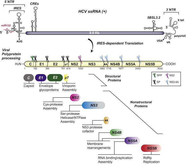
Hepatitis C virus (HCV) is a major global health burden accounting for around 170 million chronic infections worldwide. Although highly potent direct-acting antiviral drugs to treat chronic hepatitis C have been approved recently, owing to their high costs and limited availability and a large number of undiagnosed infections, the burden of disease is expected to rise in the next few years. In addition, HCV is an excellent paradigm for understanding the tight link between a pathogen and host cell pathways, most notably lipid metabolism. HCV extensively remodels intracellular membranes to establish its cytoplasmic replication factory and also usurps components of the intercellular lipid transport system for production of infectious virus particles. Here, we review the molecular mechanisms of viral replicase function, cellular pathways employed during HCV replication factory biogenesis, and viral, as well as cellular, determinants of progeny virus production.


Figures

Figure 1
Figure 1
HCV Genome Organization and Polyprotein Processing The single-strand (ss) HCV RNA genome is shown on the top. Secondary structures of cis-acting RNA elements (CREs) in the nontranslated regions (NTRs) and the coding region are schematically depicted. Interaction sites with miR-122 in the 5′ NTR that contains an internal ribosome entry site (IRES) are indicated. The polyprotein precursor and cleavage products are shown below. Numbers refer to amino acid positions of the JFH-1 isolate (GenBank accession number AB047639). Scissors indicate proteases responsible for polyprotein cleavage. SP, signal peptidase; SPP, signal peptide peptidase. Functions of cleavage products are indicated below each viral protein. RdRp, RNA-dependent RNA polymerase. VR, variable region in the 3′ NTR.
Figure 2
Figure 2
Model of an HCV-Induced Double-Membrane Vesicle and Hypothetical 3D Structures of Membrane-Associated HCV Proteins Virus-induced double-membrane vesicles (DMVs) contain HCV nonstructural proteins and RNA and are sites of active RNA replication. The DMV might contain a (transient) opening or a distinct transporter to allow exchange of nucleotides and viral RNA of the DMV interior with the cytoplasm. Note that the viral replicase might also reside on the outer surface of the DMV (not shown). Ribbon diagrams of membrane-associated HCV proteins and assumed conformational changes required for replication are indicated in boxes on the right. NS5B: the structure on the left corresponds to the closed conformation, representing the potential initiation state of the enzyme. The panel on the right shows NS5B in a hypothetical elongation mode. This conformational change would release the RNA binding groove to accommodate a dsRNA replication intermediate (shown in blue and yellow). NS5A: model of a full-length dimer associated to a membrane via the N-terminal amphipathic α helix. Only the clam-like dimer (Tellinghuisen et al., 2005) is shown for simplicity. Domains (D) 2 and 3 are intrinsically unfolded and thought to interact with multiple co-opted host factors, including cyclophilin A (CypA) binding to D2. NS3-4A complex: the presumed membrane orientation of the NS3-4A complex during polyprotein synthesis and prior to self-cleavage at the NS3/4A site is shown on the left. After cleavage, profound structural changes occur, most notably a membrane insertion of the C-terminal tail of NS4A (orange) and a repositioning of the C-terminal NS3-helicase domain (gray) away from the membrane. NS4B: the proposed dual membrane topology is shown. Structures of amphipathic α helices AH2 and H2 have been determined experimentally, while the other structural elements are based on in silico predictions. AH2 potentially traverses the membrane posttranslationally. Structure models are adapted from Bartenschlager et al. (2013) with permission from the publisher and Francois Penin.
Figure 3
Figure 3
HCV-Mediated Subversion of Lipid Homeostasis Implicated in MW Biogenesis HCV induces extensive remodeling of intracellular membranes, most notably double-membrane vesicles (DMVs) and less frequently multimembrane vesicles (MMVs). Right panel: model of HCV-subverted host cell proteins and lipids implicated in MW biogenesis. PI4KIIIα interacts with NS5A and NS5B to induce elevated levels of PI4P (1). Subsequently, oxysterol-binding protein (OSBP) delivers cholesterol to HCV-remodeled membranes. OSBP recruitment is facilitated by locally elevated PI4P levels as well as interactions with VAPs and NS5A (2). For comparison, a naive cell is shown in the left panel.
Figure 4
Figure 4
Model of HCV Particle Production Left panel: hypothetical model of HCV assembly. Viral progeny RNA is shuttled from replication sites to cytosolic lipid droplets (cLDs), facilitated by NS3 and NS5A. Core protein eventually rerecruited from cLDs to the ER is thought to trigger nucleocapsid formation and budding into the ER lumen. Presumably by tight interaction between NS2 and NS3 protease domains, NS2 brings together structural and nonstructural proteins. Nascent virions incorporate cellular lipoproteins, especially ApoE, presumably required for lipidation of virus particles. This lipidation might occur during budding, according to the hybrid particle model, or during egress via interaction between the virion and VLDL particles, according to the dual-particle model (right panel).

References

    1. Ago H., Adachi T., Yoshida A., Yamamoto M., Habuka N., Yatsunami K., Miyano M. Crystal structure of the RNA-dependent RNA polymerase of hepatitis C virus. Structure. 1999;7:1417–1426. - PubMed
    1. Altan-Bonnet N., Balla T. Phosphatidylinositol 4-kinases: hostages harnessed to build panviral replication platforms. Trends Biochem. Sci. 2012;37:293–302. - PMC - PubMed
    1. André P., Komurian-Pradel F., Deforges S., Perret M., Berland J.L., Sodoyer M., Pol S., Bréchot C., Paranhos-Baccalà G., Lotteau V. Characterization of low- and very-low-density hepatitis C virus RNA-containing particles. J. Virol. 2002;76:6919–6928. - PMC - PubMed
    1. Appel N., Zayas M., Miller S., Krijnse-Locker J., Schaller T., Friebe P., Kallis S., Engel U., Bartenschlager R. Essential role of domain III of nonstructural protein 5A for hepatitis C virus infectious particle assembly. PLoS Pathog. 2008;4:e1000035. - PMC - PubMed
    1. Appleby T.C., Anderson R., Fedorova O., Pyle A.M., Wang R., Liu X., Brendza K.M., Somoza J.R. Visualizing ATP-dependent RNA translocation by the NS3 helicase from HCV. J. Mol. Biol. 2011;405:1139–1153. - PMC - PubMed

Publication types

LinkOut - more resources